Influenza vaccination is universally recognized as the most effective measure to prevent influenza infection and transmission. Commercially available influenza vaccines are mainly classified into two categories by manufacturing technology: live attenuated influenza vaccines (LAIV) and inactivated influenza vaccines (IIV). Inactivated vaccines are further divided into split vaccines and subunit vaccines, among which influenza split vaccines dominate the market.
Influenza vaccines are manufactured via blending, filtration and filling of monovalent bulks. The production of monovalent bulks for influenza split vaccines serves as the core procedure throughout the whole manufacturing workflow, which directly determines the quality and safety of final products. Currently, industrial production of monovalent bulks for influenza split vaccines primarily adopts 9–11 day-old embryonated chicken eggs for viral propagation. The workflow encompasses allantoic fluid harvesting, purification, splitting, inactivation and filtration to obtain qualified influenza viral antigens. While Madin-Darby Canine Kidney (MDCK) cell culture is being investigated as an alternative to egg-based production, this article focuses elaborately on the manufacturing process of egg-derived influenza split vaccines.
Developed on the basis of whole-virus inactivated influenza vaccines, influenza split vaccines are produced by treating influenza viruses with optimized splitting agents under controlled conditions. This process removes viral nucleic acids and macromolecular proteins, while retaining key immunogenic components including hemagglutinin (HA), neuraminidase (NA), as well as partial matrix (M) proteins and nucleoprotein (NP). Residual splitting agents are subsequently eliminated and target antigens are further purified to yield final products. Technically, egg-based split vaccine processes are categorized into conventional and modern versions; the latter incorporates an additional purification step to further enhance antigen purity. In the late 20th century, egg-derived influenza subunit vaccines were developed from split vaccines. By targeted cleavage of viral envelope proteins HA and NA and subsequent intensive purification, final antigens are predominantly composed of HA and NA proteins.
The detailed manufacturing procedures of modern egg-based influenza split vaccines are specified as follows:
1. Viral Inoculation and Propagation
Qualified 9–11 day-old embryonated eggs with active embryonic movement and clear, evenly distributed blood vessels are selected via candling in a darkroom. Eggs are placed on egg trays with the air sac facing upward, then immersed in disinfectant-containing thermostatted water bath for 2 minutes before being transferred to the inoculation room.
Viral seeds are diluted with phosphate buffered saline (PBS). After assembling inoculation needles, supports and seed vials, air evacuation is performed under manual operation. Upon completion of degassing, the system is switched to automatic mode, and egg trays are loaded into the automatic inoculator. Post inoculation, eggs are transferred to incubators for cultivation. The incubation parameters are set as follows: H1N1 and H3N2 strains are cultured at 33.0–34.0 °C for 48–72 hours; B/Yamagata and B/Victoria strains are incubated at a constant temperature of 33.0 °C for 48–72 hours.
2. Harvest of Egg Allantoic Fluid
Eggs are re-examined by candling in a darkroom to discard dead embryos, embryos with deviated air sacs and weak embryos; only eggs with robust blood vessels are retained. Selected eggs are transferred to a cold storage chamber at 2–8 °C and cooled for no less than 16 hours, then delivered to the clean room for fluid collection.
The automatic harvester is disinfected with 75% ethanol solution, followed by installation of harvesting heads, collection tanks and shell-cutting blades. A vacuum pump is connected to the collection tank. The equipment is set to automatic mode, and egg trays are loaded to start allantoic fluid collection. Harvested allantoic fluid is either temporarily stored in cold storage or directly delivered to the next production stage.
Formaldehyde is a classic inactivating agent widely applied in biological product manufacturing. Its inactivation mechanism lies in reacting with amino-containing nucleobases of viral nucleic acids and viral envelope proteins: it induces protein denaturation and blocks the release of nucleic acids from viral particles, thereby completely eliminating viral infectivity. Formaldehyde is the predominant inactivator used by domestic manufacturers, while some overseas enterprises adopt β-propiolactone for viral inactivation.
Two mainstream inactivation-purification sequences are adopted in industrial production: pre-inactivation (inactivation prior to purification) and post-inactivation (purification prior to inactivation). Taking formaldehyde inactivation as an example, post-inactivation features better process controllability. Since formaldehyde dosage is calculated based on the volume of feed liquid, higher viral purity with fewer impurities enables formaldehyde to exert stronger inactivation efficacy at a fixed dosage. In contrast, pre-inactivation provides superior microbial control throughout the workflow.
The standard operating procedure for post-inactivation using formaldehyde is described below: 0.2% formaldehyde solution is added into monovalent viral split fluid at a volume ratio of 5%±0.5% for influenza A strains and 2.5%±0.25% for influenza B strains, to reach a final formaldehyde concentration of approximately 0.01% (W/V, 100 μg/mL) for type A strains and 0.005% (W/V, 50 μg/mL) for type B strains. The mixture is stirred continuously for no less than 15 minutes. Inactivation is carried out at 15–25 °C with intermittent stirring for 25–26 hours, followed by static storage at 2–8 °C for 24–96 hours.
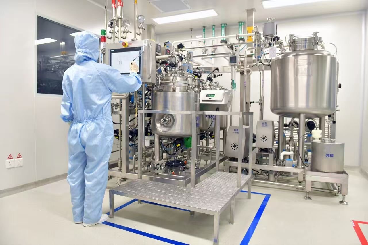
4. Clarification and Ultrafiltration Concentration
Crude allantoic fluid is first filtered through filter cloth to remove insoluble contaminants such as eggshell debris, then clarified via continuous-flow centrifugation at 8000 rpm to eliminate macromolecular substances and red blood cells. The clarified supernatant is concentrated via tangential flow ultrafiltration using membrane cassettes with molecular weight cut-off (MWCO) ranging from 300 kDa to 1000 kDa. The transmembrane pressure is maintained at 1.2 bar, and the pressure differential between the inlet and outlet of membrane cassettes is controlled below 0.7 bar to obtain concentrated viral fluid. Sampling is conducted to detect bacterial endotoxin, HA content and total protein content.
Clarification aims to remove particulate matters in allantoic fluid. Manufacturers adopt diverse strategies including high-speed continuous-flow centrifugation, direct filtration, or combined filtration and centrifugation, to reduce microbial load and eliminate yolk residues, so as to improve the operating efficiency of subsequent ultrafiltration. Tangential flow ultrafiltration is universally applied to reduce liquid volume and elevate antigen concentration: viral particles are retained and recycled via the retentate stream, while small-molecule impurities such as urate permeate through the membrane and are discarded. The selection of ultrafiltration membrane MWCO (100 kDa, 300 kDa, 500 kDa, 750 kDa or 1000 kDa) depends on the downstream purification capacity of individual production lines.
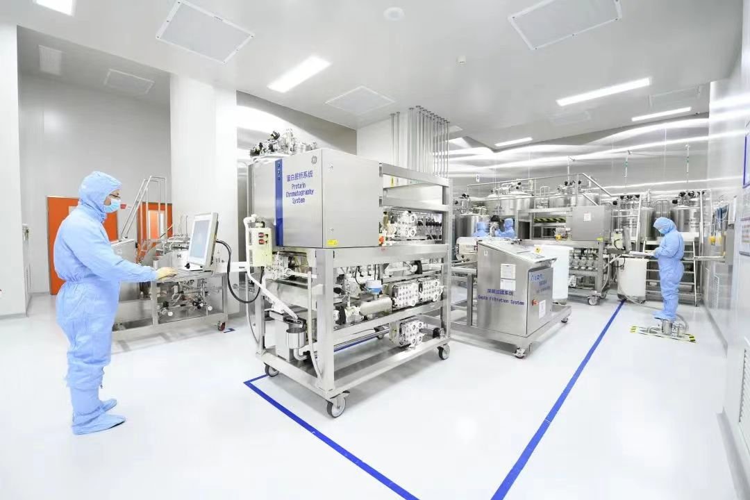
5. Purification (Centrifugation or Chromatography)
Major purification technologies for influenza viruses include ultrafiltration, ultracentrifugation, gel filtration chromatography and mixed-mode chromatography.
Ultrafiltration: A membrane separation technology that selectively separates and recovers target substances via microporous structures of semipermeable membranes under external pressure (0.1–0.5 MPa).
Ultracentrifugation: A highly efficient method for separating subcellular components and protein macromolecules, including differential centrifugation and density gradient centrifugation. Differential centrifugation separates particles with distinct molecular sizes through alternating low and high-speed centrifugation. Density gradient centrifugation achieves separation based on differences in sedimentation rate under ultrahigh centrifugal force, with sucrose, glycerol and Ficoll as common density media. Sucrose density gradient centrifugation is the most widely adopted approach for influenza virus purification.
Mixed-mode chromatography: Media with complex structures integrating multiple chromatographic functions (e.g., hydrophobic interaction and ion exchange chromatography).
A two-step purification process is generally implemented for concentrated viral fluid to enhance product purity and reduce ovalbumin content.
Centrifugal purification: The concentrated sample is centrifuged at 35000 rpm. Influenza viruses distributed within the 35%–45% sucrose gradient fraction are collected as purified viral fluid.
Chromatographic purification: Sucrose is removed from centrifuged viral fluid via ultrafiltration. After dilution, the sample is loaded onto gel filtration columns. The first peak detected by ultraviolet monitoring represents the target viral fraction, which is collected as purified antigen.
Purification strategies vary among manufacturers. International pharmaceutical enterprises including Sanofi Pasteur, GSK and Novartis, as well as research institutions, predominantly adopt sucrose density gradient centrifugation for whole-virus purification. Domestic manufacturers apply gel chromatography, sucrose density gradient centrifugation, or a combination of both methods to guarantee vaccine quality and immunogenicity.
Comparative analysis shows that gel chromatography has prominent limitations for large-scale production: high viscosity of concentrated allantoic fluid leads to frequent column cleaning, repacking and performance verification, resulting in significant batch-to-batch variation. The process consumes large volumes of PBS, alkaline solutions and other buffers, with increased risk of contamination. In addition, gel media have short service life and raise production costs. By contrast, sucrose density gradient centrifugation features simple workflows, consistent batch quality, high yield and purity, low ovalbumin residue, low production cost and superior safety profile, making it more suitable for large-scale commercial manufacturing.
Viral splitting effectively alleviates adverse reactions after vaccination while preserving high immunogenicity. A variety of splitting agents are available for influenza virus processing, such as diethyl ether, sodium deoxycholate, Triton X-100, Triton N-101, n-octyl-β-D-glucopyranoside and cetyltrimethylammonium bromide (CTAB). Single or combined splitting agents are applied in production, among which Triton X-100 is the most commonly used. For instance, viral splitting can be performed at room temperature for 60 minutes using Triton X-100 at a final concentration of 0.5%.
Residual splitting agents and inactivators must be thoroughly removed to ensure vaccine safety and efficacy. Ultrafiltration is adopted to eliminate residual Triton X-100 and free formaldehyde.
The processed antigen solution undergoes sterile filtration through a combined filter cartridge with pore sizes of 0.45 μm and 0.2 μm. The filtrate is diluted 1.5–2 times with PBS to prepare monovalent bulks of influenza split vaccine.
9. Formulation of Semi-finished Products
Trivalent influenza vaccines contain three influenza strains. After HA quantification of three monovalent bulks, semi-finished products are formulated with the HA concentration of each strain controlled within 30–36 μg/mL. The same formulation principle applies to quadrivalent influenza vaccines.
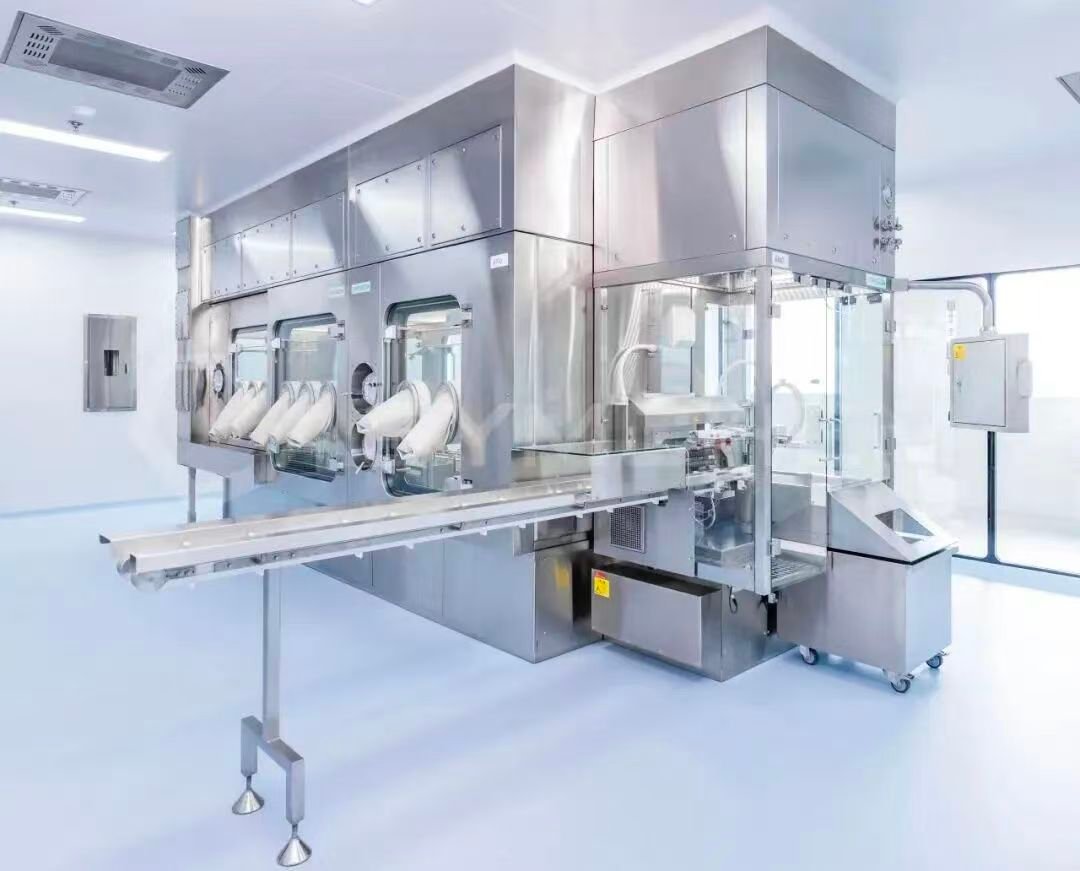
Finished vaccines are packaged in two forms: vials and pre-filled syringes. Semi-finished products are filled into final containers and subjected to comprehensive quality testing. Products passing all quality specifications are released as qualified finished vaccines.
Conventional influenza split vaccines are subviral vaccines derived from whole inactivated viruses via splitting treatment, consisting of surface antigens (HA and NA) and trace viral nucleocapsid and matrix proteins. With technological advancement, manufacturers have upgraded traditional processes by adding an extra purification step to develop modern split vaccine production lines. The resulting bulks contain highly purified HA and NA with minimal residual nucleocapsid proteins.
Egg-derived influenza subunit vaccines share a three-step purification workflow with modern split vaccines. This type of vaccine is manufactured by specific cleavage and extraction of viral envelope proteins HA and NA, featuring higher purity of HA but lower NA content compared with split vaccines. Nevertheless, subunit vaccines exhibit relatively weak immunogenicity, so adjuvants are required to achieve satisfactory immune effects.
In terms of stability, egg-derived subunit vaccines are slightly inferior to split vaccines: they are more sensitive to temperature fluctuation and have a shorter shelf life, while both products comply with the same storage requirement (2–8 °C refrigeration). Such differences are determined by antigen composition and structural characteristics. Subunit vaccines reduce adverse reactions by simplifying antigen components at the cost of decreased stability; split vaccines retain structures closer to native viruses, thus maintaining strong immunogenicity and better environmental tolerance.