Cell culture medium (CCM) components affect cell growth, as well as the production and critical quality attributes (CQAs) of monoclonal antibodies (mAbs) and recombinant proteins. Iron is an essential constituent of CCM and participates in numerous cellular processes. Nevertheless, iron triggers reactive oxygen species (ROS) generation via the Fenton reaction, which further induces cellular damage. Accordingly, iron ion concentration exerts a pronounced impact on antibody quality.
This article elaborates on the effects of iron ions on the growth of Chinese hamster ovary (CHO) cells and product quality through experimental cases.
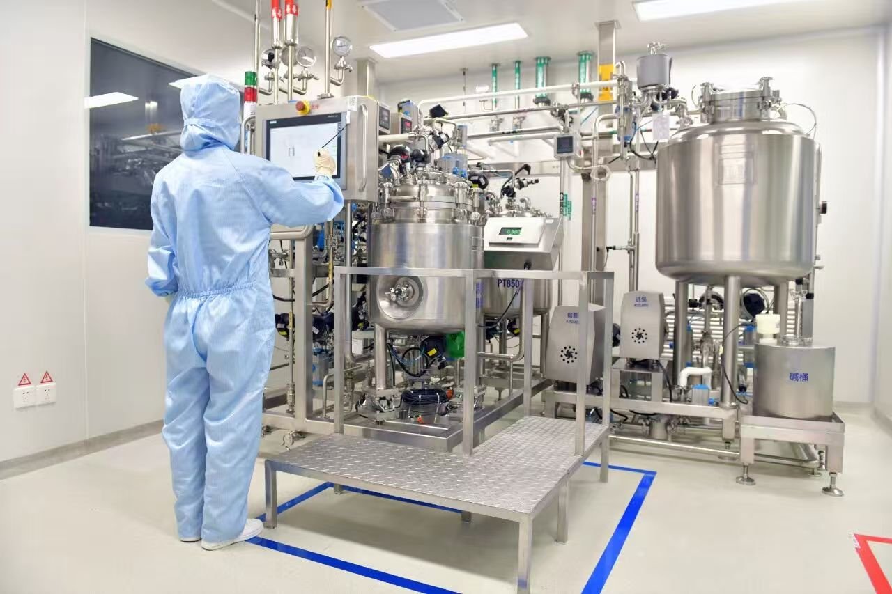
CHO cell culture processes are widely applied for the production of therapeutic antibodies and recombinant proteins targeting tumors, hematological disorders and other diseases. Maintaining stable cell performance and final product quality is a core objective for bioprocess development, which can be influenced by cell lines, process parameters and CCM compositions.
Major CCM ingredients include amino acids, carbohydrates, vitamins, lipids, inorganic salts and trace elements. Each component substantially modulates cell performance and CQAs of end products. Therefore, it is critical to clarify their functions in cellular metabolism and protein expression. As an indispensable element for cellular activities, iron acts as a cofactor for a variety of enzymes involved in energy metabolism, DNA biosynthesis and repair, as well as antioxidant defense systems.
Given the significant influence of iron ions in culture media and additives on cell growth and product quality, exploring the correlation between iron ion concentration and product yield & quality is of great practical significance.
Cell line: CHO-K1
Tested iron supplements: Ammonium ferric citrate (FAC), Ferric citrate (FC)
Cultivation protocol: Cultures were performed in 50 mL TPP tubes. The initial seeding density was 2×10⁵ cells/mL. Cultivation was carried out at 37 °C with 5% CO₂, at a working volume of 30 mL for a total duration of 17 days. Samples were collected daily to determine cell density, cell viability and biochemical parameters.
3.1 Effects of elevated FAC concentration on cell performance and IgG quality attributes
As shown in Figure 1, FAC concentration significantly affected cell performance. Compared with the group supplemented with 2 mg/L FAC, groups with 10 mg/L, 50 mg/L and 100 mg/L FAC achieved higher peak viable cell density (peak VCD). The antibody titer also increased within the first 16 days of cultivation. However, cell viability declined sharply from Day 12 onwards. The group with 2 mg/L FAC maintained favorable cell status and obtained the highest product titer on Day 17.
As presented in Figure 2, elevated FAC concentration caused a dose-dependent increase in high molecular weight (HMW) aggregates, rising from 0.9% (2 mg/L FAC) to 2.8% (100 mg/L FAC), which was negatively correlated with the percentage of main peak. Iron concentration also exerted a prominent dose-dependent effect on protein glycosylation.
When FAC concentration increased from 2 mg/L to 100 mg/L, the level of terminal galactosylation increased from 13.3% to 25.6%, accompanied by a reduction in terminal N-acetylglucosamine (GlcNAc) content. For mAb1, the levels of terminal sialylation, mannosylation and unidentified glycan species were below 0.7%, 1.4% and 1.2% respectively (Figure 2b), and remained unaffected by iron concentration.
In summary, increased FAC concentration elevated peak VCD but impaired long-term cell viability. Meanwhile, it slightly raised HMW aggregate content and enhanced terminal galactosylation of mAb1.
3.2 Effects of different iron sources on cell performance and IgG quality attributes
Replacing FAC with FC as the iron source led to inhibited cell growth. At concentrations of 10 mg/L, 50 mg/L and 100 mg/L, the peak VCD of FC groups decreased by 7.4%, 28.9% and 3.5% respectively compared with the corresponding FAC groups.
For both iron sources, higher iron concentration accelerated the drop of cell viability. Moreover, cell viability declined more rapidly in all FC groups than in the counterpart FAC groups at the same concentration. Only marginal differences in antibody titer curves were observed between the two iron sources (Figure 3c). Overall, regardless of iron dosage, FC resulted in faster declines of VCD and cell viability than FAC, indicating inherent differences between the two iron sources.
Distinct glycosylation profiles were also observed between the two iron sources. Increased FAC promoted terminal galactosylation (13.3% at 2 mg/L up to 25.6% at 100 mg/L). In contrast, FC reduced terminal galactosylation from 11.5% (2 mg/L) to 8.0% (50 mg/L), showing a negative correlation with terminal GlcNAc level. These results demonstrated that the glycosylation discrepancy between FAC and FC was independent of iron concentration, and was attributed to other unknown properties of the two iron sources.
3.3 Analysis of trace element impurities in FAC and FC
Certain elements including magnesium (Mg), aluminum (Al), calcium (Ca), titanium (Ti), vanadium (V), cobalt (Co), copper (Cu), zinc (Zn) and gallium (Ga) were only detected in FAC. By contrast, boron (B) and potassium (K) had higher contents in FC and were undetectable in FAC. Chromium (Cr), manganese (Mn) and nickel (Ni) were present at higher levels in FAC than in FC.
Manganese is a well-documented regulator of antibody galactosylation. Impurity-derived manganese from FAC accounted for over 94% of the total manganese in the culture medium, suggesting that manganese was a highly relevant interfering factor.
3.4 Effects of iron sources with high and low manganese impurities on cell performance and IgG quality attributes
Two types of FC were used in this study: commercially sourced FC with high manganese impurities (FC<sub>Purch</sub>) and synthetic FC with low manganese impurities (FC<sub>Synt</sub>), at test concentrations of 10 mg/L, 50 mg/L and 100 mg/L.
Regardless of iron concentration, FC<sub>Purch</sub> supported better cell growth than FC<sub>Synt</sub>. Supplementing extra manganese into FC<sub>Synt</sub> restored cell VCD to a comparable level. Cells cultured with FC<sub>Purch</sub> or manganese-supplemented FC<sub>Synt</sub> maintained viability for a longer period relative to the original FC<sub>Synt</sub> groups.
IgG titers in groups with FC<sub>Purch</sub> and manganese-supplemented FC<sub>Synt</sub> were higher than those in pure FC<sub>Synt</sub> groups, proving that manganese improved specific productivity. The content of HMW aggregates of mAb1 was below 5.5% across all test groups, with absolute differences less than 3.3% at the same iron concentration. Additionally, manganese supplementation increased the level of terminal galactosylation.
Iron and its associated impurities jointly affect cell culture performance and glycosylation of recombinant proteins. Manganese, as a common impurity in iron raw materials, can improve overall cell culture performance, whereas excessive iron impairs cell growth, viability and antibody titer. Furthermore, iron and manganese exert opposing effects on the glycosylation of the target recombinant protein.
It is essential to adopt iron sources with low impurity content to eliminate the interference of iron and related impurities in cell culture. Rational control of the concentration of individual elements enables the establishment of a robust and consistent cell culture process, and ensures excellent reproducibility of CQAs for recombinant biotherapeutics.