


With the rapid advancement of precision medicine and nucleic acid therapeutics, lipid nanoparticles (LNPs) and liposomes have emerged as pivotal delivery nanocarriers, underpinning the research and industrialization of mRNA vaccines, siRNA therapeutics and a broad spectrum of biomacromolecular drugs. The therapeutic efficacy of such nanocarriers is fundamentally determined by the precise modulation of their physicochemical profiles. Key parametric indicators, including lipid composition, particle size, polydispersity index (PDI), zeta potential, morphological microstructure and encapsulation efficiency (EE%), collectively dictate formulation stability, in vivo biodistribution and cellular internalization capacity. Based on standardized characterization practices and global regulatory guidelines, this review systematically elaborates the critical quality attributes (CQAs), key influencing factors and state-of-the-art analytical techniques of LNPs and liposomes, aiming to provide a rigorous technical reference for pharmaceutical development.
1. Regulatory Status of LNP Products
To date, no universal global regulatory framework has been established to cover all categories of LNP-based formulations. Current regulatory requirements are predominantly tailored to payload categories and clinical indications. Regulatory authorities including the CDE, EMA, FDA and WHO have issued targeted guidance for mRNA-LNP vaccines, whereas specialized regulatory specifications for therapeutic RNA-LNP formulations remain in continuous iteration and optimization. In addition, the core focus of biodistribution assessment differs significantly between vaccine and therapeutic LNP applications. Accordingly, preclinical developers are recommended to conduct proactive regulatory communication prior to initiating formal investigative studies.
In accordance with the FDA guidance Liposome Drug Products: Chemistry, Manufacturing, and Controls; Human Pharmacokinetics and Bioavailability; and Labeling Documentation, the quality characterization of organic nanoparticle formulations prioritizes physicochemical CQAs such as particle size, size distribution and microscopic morphology. Deviations in these critical parameters may lead to compromised delivery potency, increased payload leakage and inferior batch-to-batch consistency, ultimately resulting in inconsistent clinical performance and therapeutic failure.
2. Critical Quality Attributes (CQAs) of LNPs
2.1 Chemical Composition Profiling
LNPs are fabricated via spontaneous self-assembly, a process fundamentally distinct from conventional top-down chemical synthesis. The self-assembly mechanism is extremely sensitive to manufacturing parameters, wherein subtle process variations can induce profound alterations in nanoparticle physicochemical properties.
A canonical LNP formulation consists of four major lipid components with fixed molar ratios:
Ionizable/cationic lipids (approximately 50 mol%): neutrally charged under physiological pH conditions and positively charged in acidic microenvironments, enabling efficient nucleic acid encapsulation and endosomal escape to facilitate intracellular delivery.
Helper lipids (e.g., DSPC, approximately 10 mol%): constitute the basic skeleton of lipid bilayers and maintain structural integrity of nanoparticles.
Sterol lipids (predominantly cholesterol): modulate the rigidity, compactness and structural stability of lipid membranes.
PEGylated lipids (≤ 3 mol%): anchor on the particle surface, serving as key modulators of particle size, colloidal stability, immune evasion capability and payload encapsulation performance.
Precise stoichiometric control of lipid components is indispensable for guaranteeing formulation stability and delivery efficacy. For instance, the depletion of PEGylated lipids will trigger abnormal particle enlargement (typically >200 nm), elevated polydispersity and enhanced immune recognition, which severely impair the targeted delivery capacity of LNPs. LNP component ratios are primarily determined by formulation design. For quantitative verification, ultra-high performance liquid chromatography (UHPLC) coupled with a charged aerosol detector (CAD) is widely adopted, with quantitative results expressed as mass concentration (mg/mL) and molar percentage of individual components.
YaoHai Bio has established a validated UHPLC-CAD quantitative method for LNP formulations (ALC-0315 system). The method achieves complete baseline separation of four lipid components, enabling accurate identification and precise quantification of all formulation constituents. All calibration curves exhibit excellent linearity with coefficient of determination (R²) values greater than 0.99, confirming superior accuracy and reproducibility for routine quality testing.
2.2 Encapsulation Efficiency (EE%)
EE% is defined as the mass ratio of encapsulated mRNA payload to the total mRNA content (sum of encapsulated and free mRNA) in LNP formulations. Optimized mRNA-LNP systems can achieve an EE% exceeding 90%, whereas liposomal formulations loaded with inorganic nanoparticles (e.g., gold nanoparticles) generally exhibit lower encapsulation levels.
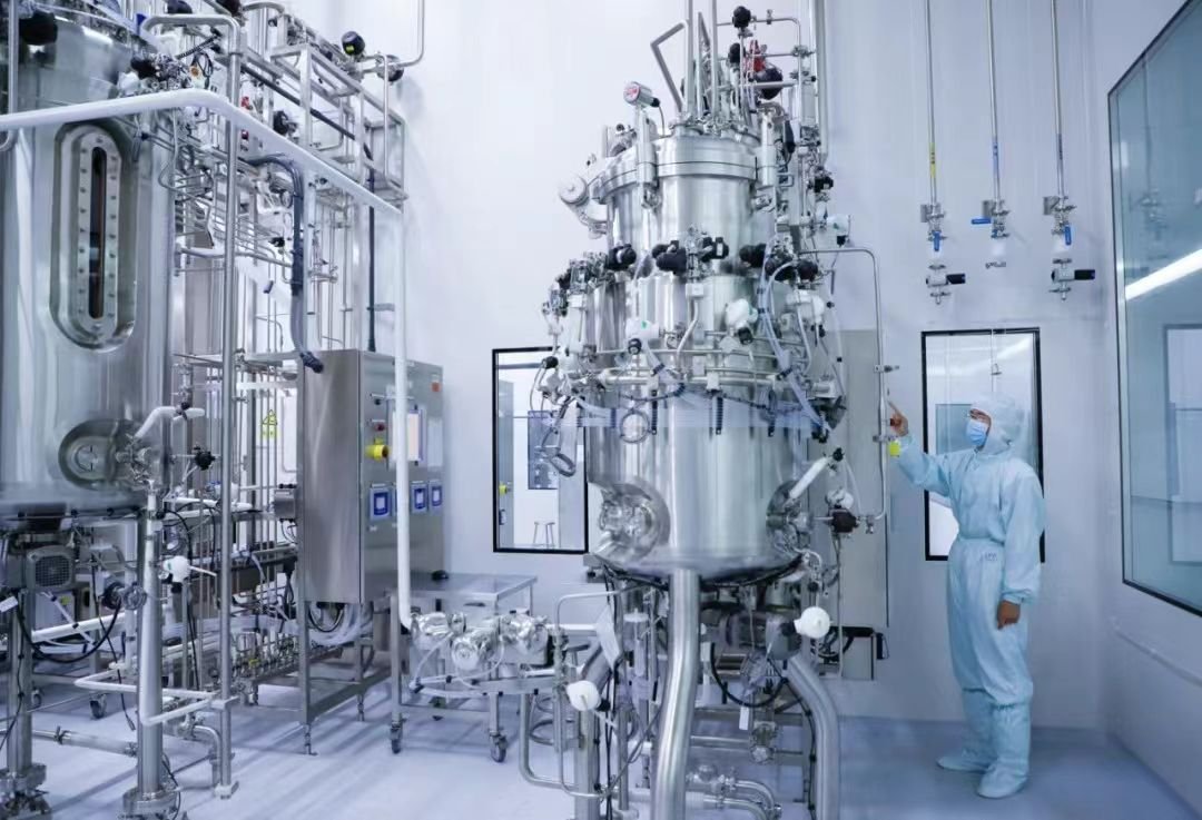
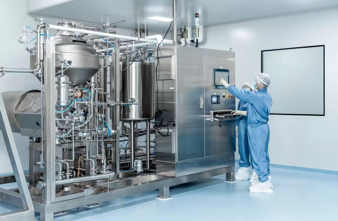
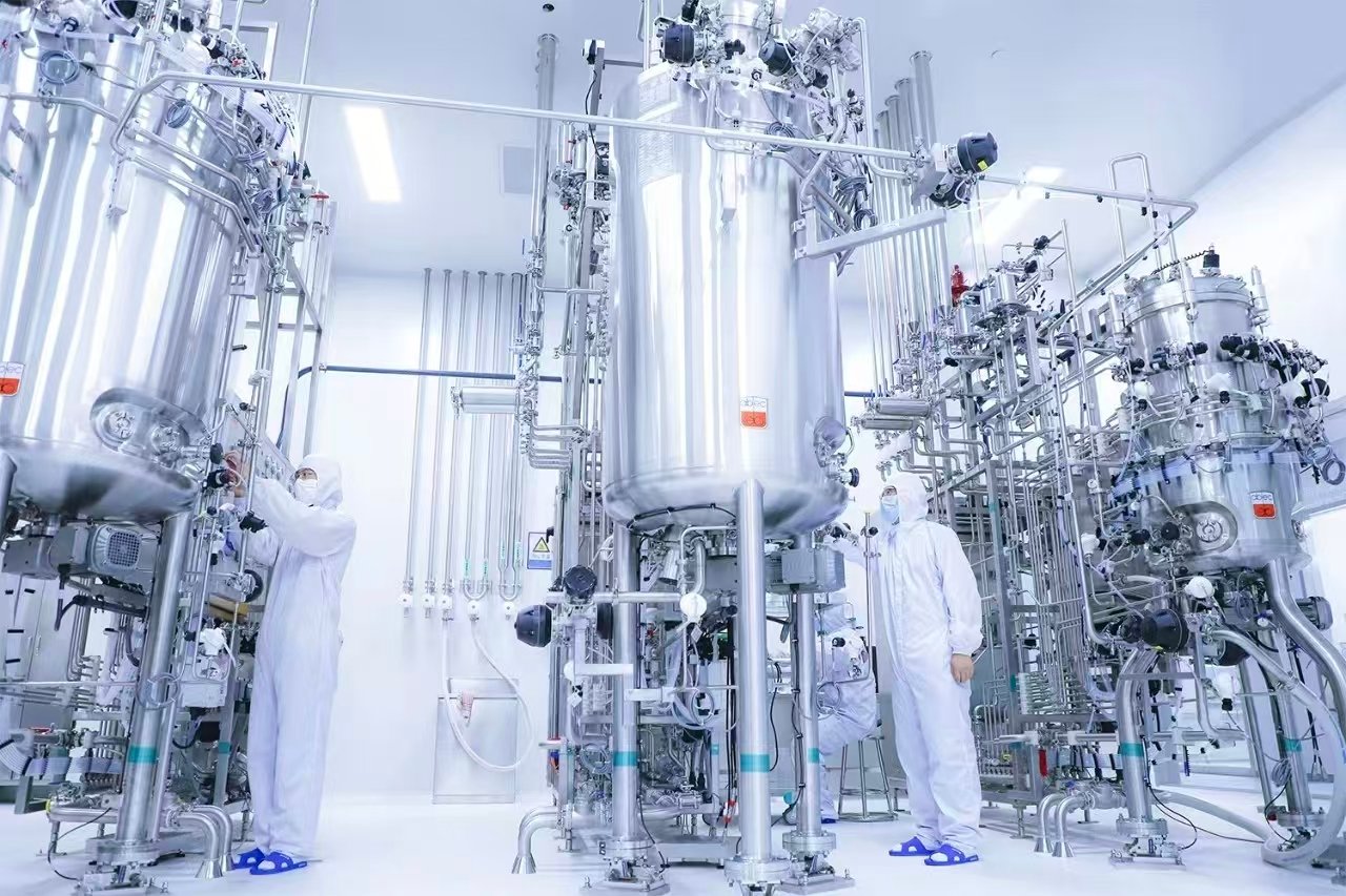
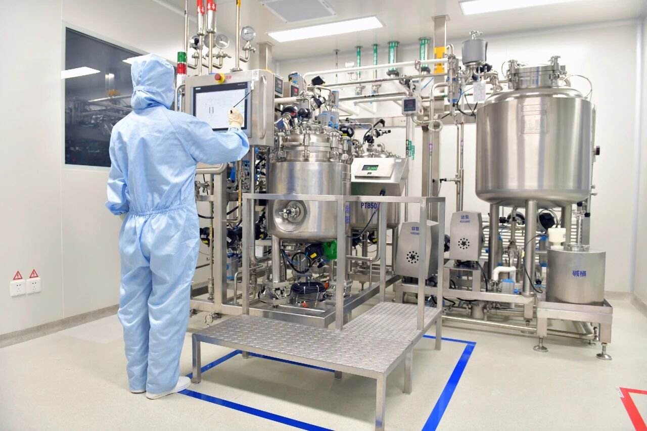
Manufacturing technology represents the dominant factor affecting LNP encapsulation performance. For identical formula compositions and payloads, different preparation strategies (manual blending, vortex mixing, microfluidic assembly) can lead to more than two-fold differences in final EE values. Microfluidic technology, characterized by highly controllable reaction conditions, currently delivers the highest encapsulation efficiency and optimal batch-to-batch reproducibility, serving as the mainstream high-standard preparation platform.
The quantitative paradigm for EE determination relies on the separation and differential quantification of free and encapsulated payloads. For RNA-loaded LNPs, fluorescence spectroscopy coupled with RiboGreen staining is the prevailing analytical method due to its high sensitivity and superior resolution.
In the standardized testing workflow established by YaoHai Bio, free RNA and total RNA concentrations span distinct order-of-magnitude ranges. Separate high-precision calibration curves are established for the two analytes with qualified R² values. Demulsified encapsulated RNA samples and free RNA samples are diluted proportionally to fit the linear range of calibration curves, thereby ensuring accurate and reliable EE% calculation results.
2.3 Physicochemical Property Characterization
Physicochemical properties are core CQAs that govern the colloidal stability, cellular uptake efficiency and in vivo biodistribution of LNP formulations. Any abnormal fluctuation in these parameters may directly reduce transfection potency, induce payload leakage and compromise therapeutic safety and efficacy.
2.3.1 Particle Size
Particle size is a paramount quality attribute, which requires targeted optimization based on administration routes and cellular targeting requirements. Excessively small nanoparticles are prone to renal clearance and potential toxicological risks, while oversized particles exhibit poor cellular internalization efficiency. Conventionally, the viable particle size range of LNPs is 50–200 nm, with the optimal therapeutic window concentrated at 80–100 nm. For tumor-targeting delivery scenarios, smaller particle sizes (approximately 60 nm) are often preferred. Ligand-modified targeted LNPs, as a cutting-edge delivery system, generally maintain a particle size of 100–160 nm.
Particle size is primarily modulated by lipid composition (especially the proportion of PEGylated lipids) and manufacturing processes. Existing studies have verified that PEG-lipid-deficient LNPs present increased particle diameter, higher polydispersity and deteriorated colloidal stability. In contrast, payload types (siRNA vs. mRNA) exert negligible effects on final particle size. Among all preparation techniques, microfluidic assembly features precise process control and exceptional batch consistency, and has been recognized as the gold-standard manufacturing strategy for uniform-sized LNPs.
Commonly adopted particle sizing techniques are summarized as follows:
Dynamic Light Scattering (DLS): enables rapid batch screening and comparative analysis, with limitations in absolute quantitative accuracy.
Multi-Angle Dynamic Light Scattering (MADLS): provides enhanced resolution and enables identification of heterogeneous particle subpopulations.
Tunable Resistive Pulse Sensing (TRPS): achieves synchronous detection of particle size and absolute particle concentration.
Nanoparticle Tracking Analysis (NTA): characterizes particle motion based on Brownian diffusion, with inferior detection efficiency and reproducibility compared with DLS.
Transmission Electron Microscopy (TEM) & Cryogenic Transmission Electron Microscopy (Cryo-TEM): enables direct visualization of particle dimension and internal microstructure (Cryo-TEM exclusive), though with low testing throughput and high instrumental and operational requirements.
In industrial and academic practice, orthogonal characterization combining DLS and electron microscopy is universally implemented to eliminate methodological errors and improve data reliability.
2.3.2 Polydispersity Index (PDI)
PDI is defined as the ratio of the squared standard deviation of particle size distribution to the mean particle size, which quantifies the homogeneity of nanoparticle populations. A PDI value close to zero indicates a highly homogeneous formulation system, while elevated PDI reflects significant particle heterogeneity. In accordance with FDA regulatory guidelines, the acceptable PDI threshold is below 0.3; the industrial internal control specification is strictly maintained at 0.05–0.2 to reserve sufficient process tolerance.
Lipid formulation composition and manufacturing processes are the dominant influencing factors of PDI. Traditional preparation methods including thin-film hydration and high-pressure homogenization typically yield PDI values above 0.5 with poor size uniformity. In comparison, microfluidic fabrication generates LNPs with narrow size distribution and PDI values consistently below 0.2. The analytical techniques for PDI are consistent with particle sizing, wherein bulk detection methods (DLS, TRPS) are optimal for overall population homogeneity evaluation.
2.3.3 Zeta Potential
Zeta potential characterizes the effective surface charge of nanoparticles, serving as a critical indicator of colloidal suspension stability and biotoxicity risk. An absolute zeta potential value of |±30 mV| and above indicates favorable colloidal stability, while near-neutral surface charge status tends to induce particle aggregation and fusion. Excessively positive surface charge will significantly increase cytotoxicity. Hence, the ideal zeta potential range for most nanocarriers is controlled around ±30 mV.
Notably, LNPs incorporate PEGylated stabilizers to maintain colloidal stability under low-charge conditions. The industrial quality specification for LNP zeta potential is strictly controlled within |±20 mV| (near-neutral range).
Zeta potential is predominantly measured via electrophoretic light scattering (principally coupled with DLS systems) and TRPS (capable of single-particle-level zeta potential and size synchronous detection). The core influencing factors include lipid formulation and suspension pH. A decrease in environmental pH will increase surface positive charge and correspondingly elevate zeta potential values.
2.3.4 Morphology and Internal Microstructure
Nanoparticle morphology and internal architecture directly determine payload encapsulation capacity and in vivo delivery behavior. Liposomes can form unilamellar, multilamellar (onion-like) and other layered structures, wherein multilamellar vesicles enable co-encapsulation of payloads with diverse physicochemical properties and molecular sizes. The internal microstructure of LNPs is comprehensively modulated by lipid composition, payload properties and manufacturing protocols.
Given the soft-material characteristics and suspension state of LNPs, conventional bulk analytical methods fail to provide morphological and structural information. Single-particle high-resolution characterization techniques are mandatory. Cryo-TEM serves as the gold-standard approach: rapid plunge freezing in liquid nitrogen eliminates ice crystal artifacts, enabling high-resolution visualization of nanoparticle surface topography and internal nanostructure.
3. Conclusion
Targeted delivery design has evolved into the core technological pillar of modern LNP-based RNA drug development. In the future, the synergistic innovation of LNP targeted modification strategies and RNA bioengineering is expected to break the liver-targeting limitation of current RNA therapeutics, facilitating the transformation of RNA therapy into a multi-organ, systemic and precise clinical treatment platform.