Antibody drugs are vital biological agents used for the treatment of a wide range of diseases such as tumors and autoimmune disorders. Their manufacturing process comprises multiple core stages, including cell line selection and construction, culture medium and bioreactor selection, cell cultivation, antibody expression and purification, as well as antibody drug formulation. This article elaborates on the key technical procedures and workflows for antibody drug production.
Since the launch of Anti-human T Lymphocyte CD3 Murine Monoclonal Antibody, China’s first domestic antibody drug, in 1999, dozens of subsequent antibody products were predominantly imported medicines for a long period. Nevertheless, the domestic antibody industry has witnessed explosive growth over the past five years with remarkably enhanced innovation capacity. For decades, the upstream raw materials, production equipment and other critical supplies of the antibody drug industrial chain have been heavily reliant on imports. Domestic manufacturers have strived for technological breakthroughs and achieved domestic substitution in multiple fields including analytical instruments, consumables, cell culture equipment and culture media, thereby driving R&D and innovation in antibody therapeutics.
1. Cell Line Selection and Construction
Mammalian cell culture serves as the starting point for antibody drug production, which generates target antibodies via cultivated cell lines. Mammalian cell systems are the predominant expression platforms for clinically approved antibody products. Therapeutic antibodies require correct protein folding and post-translational modifications to retain biological activity, which makes mammalian cells the preferred host cells. The commonly used cell lines include Sp2/0 myeloma cells, NS0 murine myeloma cells, human embryonic kidney (HEK293) cells and Chinese Hamster Ovary (CHO) cells, among which CHO cells are the most widely applied.
CHO expression systems possess distinct advantages over other expression platforms:
Superior adaptability to suspension culture, meeting the requirements for large-scale industrial production of recombinant proteins;
The expressed antibodies share high similarity with natural antibodies in structure and function;
Extremely low risk of endogenous human virus contamination;
Stable integration of exogenous genes into the CHO cell genome;
As fibroblast cells, CHO cells barely secrete endogenous proteins, which greatly facilitates the isolation and purification of target recombinant antibodies.
The development of antibody drugs is an extremely complex process, and the construction of high-yield cell lines suitable for industrial manufacturing stands out as one of the most critical links, directly determining production efficiency and final product quality. Therefore, research on the construction and optimization of stable recombinant cell lines expressing exogenous genes is of great significance. Unlike synthetic drugs, protein therapeutics are generally produced by prokaryotic or eukaryotic expression systems, such as Escherichia coli, mammalian and non-mammalian cell lines, insect cell lines, and cell-free expression systems that have become a research hotspot in recent years.
The development of engineered cell lines — from screening high-producing mammalian cell clones to establishing master and working cell banks for clinical trials and commercial production — is a tedious and lengthy process. In accordance with conventional screening workflows, the whole procedure from cell transfection to mini-pool selection and subsequent single-clone screening typically takes 3 to 6 months. Additional serial passaging tests are conducted to verify cell line stability. After obtaining high-yield single clones, in-depth optimization and characterization are carried out during upstream cell culture process development. Overall, the entire cycle from cell line screening to the completion of cell culture process development generally lasts 6 to 12 months or even longer, varying by the R&D capabilities of individual enterprises. Although cell line construction accounts for a small proportion of total production costs, it plays an irreplaceable role in maximizing antibody yields in large-scale manufacturing. Currently, the majority of commercial antibody drugs are produced using CHO cells, owing to their capability to conduct proper protein folding, assembly and human-like post-translational modifications, making them the mainstream host cells for recombinant protein therapeutics.
2. Selection of Culture Media and Bioreactors
Clone screening during cell line development is normally performed based on a standardized cell culture platform. For pilot-scale production and Investigational New Drug (IND) applications, 2 to 3 clones from distinct lineages are usually selected, with 1 or 2 designated as backup candidates. Cell culture processes exert a profound impact on the yield, quality and safety of biological products, and the selection of cell culture media is particularly crucial. Customized culture systems, including basal media and feed supplements, are formulated to support cell growth and antibody expression. Common commercial media include DMEM and RPMI 1640.
In the screening of basal and feed media for fed-batch culture, it is recommended to match basal media with feed supplements from the same brand in the initial stage. After primary screening, culture media are categorized by functional characteristics, such as growth-promoting, viability-maintaining and expression-enhancing media. Medium combinations are optimized by blending different functional types, and Design of Experiment (DOE) methodologies are adopted for systematic process optimization.
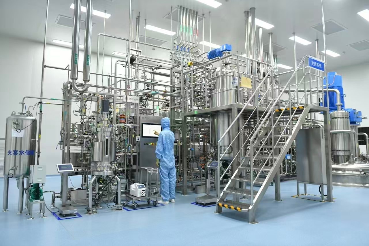
Leading biopharmaceutical enterprises such as Amgen and Genentech have achieved a protein titer of 5–10 g/L using proprietary cell lines, while certain cell lines developed by Pfizer and MedImmune even exceed 10 g/L. Correspondingly, bioreactor configurations have evolved from traditional large-scale 20 kL stainless steel vessels toward small-volume (1 kL), continuous and single-use formats. Conventional stainless steel bioreactors require rigorous cleaning and sterilization before use and carry a higher contamination risk. In contrast, single-use bioreactors drastically reduce preparation time. Stirred-tank bag bioreactors, including Hyclone Single-Use Bioreactors (SUB), Sartorius BIOSTAT and Xcellerex XDR-DSTB, are increasingly deployed in therapeutic antibody manufacturing.
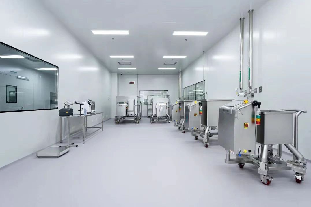
3. Cell Thawing, Subcultivation, Expansion and Large-Scale Cultivation
Taking the commercial production workflow of monoclonal antibodies (mAbs) as an example, the upstream cell culture process follows the sequence: cell thawing and subcultivation → cell expansion → large-scale cultivation → cell separation (the last step is also classified as part of downstream processing).
3.1 Cell Thawing and Subcultivation
Vials of working cell bank are placed in a thermostatted water bath at approximately 35 °C and pre-warmed for 30 minutes. Thawed cells are transferred into prepared culture media, harvested by centrifugation, and inoculated into shake flasks (e.g., 250 mL) for initial subcultivation. Key parameters including CO₂ concentration, incubation temperature, culture duration and seeding density are strictly controlled to maintain high cell viability.
Subsequently, seed cells are diluted and transferred into larger shake flasks (500 mL to 2000 mL). Critical culture parameters such as temperature, CO₂ concentration, dissolved oxygen (DO), agitation speed and pH are precisely regulated to expand cell populations until the cell density meets the inoculation criteria for the first-stage seed bioreactor.
3.2 Cell Expansion (Seed Cultivation)
Primary seed culture: Shake flask-derived seed cells are inoculated into 20 L first-stage seed bioreactors. Culture conditions including temperature, DO, agitation speed and pH are monitored and controlled to amplify cell density for inoculation into secondary seed bioreactors.
Secondary seed culture: Cells from primary seed bioreactors are transferred into 50 L secondary seed bioreactors under controlled culture parameters for further expansion to meet the inoculation requirements of tertiary seed bioreactors.
Tertiary seed culture: Cells are inoculated into 100 L tertiary seed bioreactors and cultivated until sufficient cell density is achieved for large-scale production bioreactors.
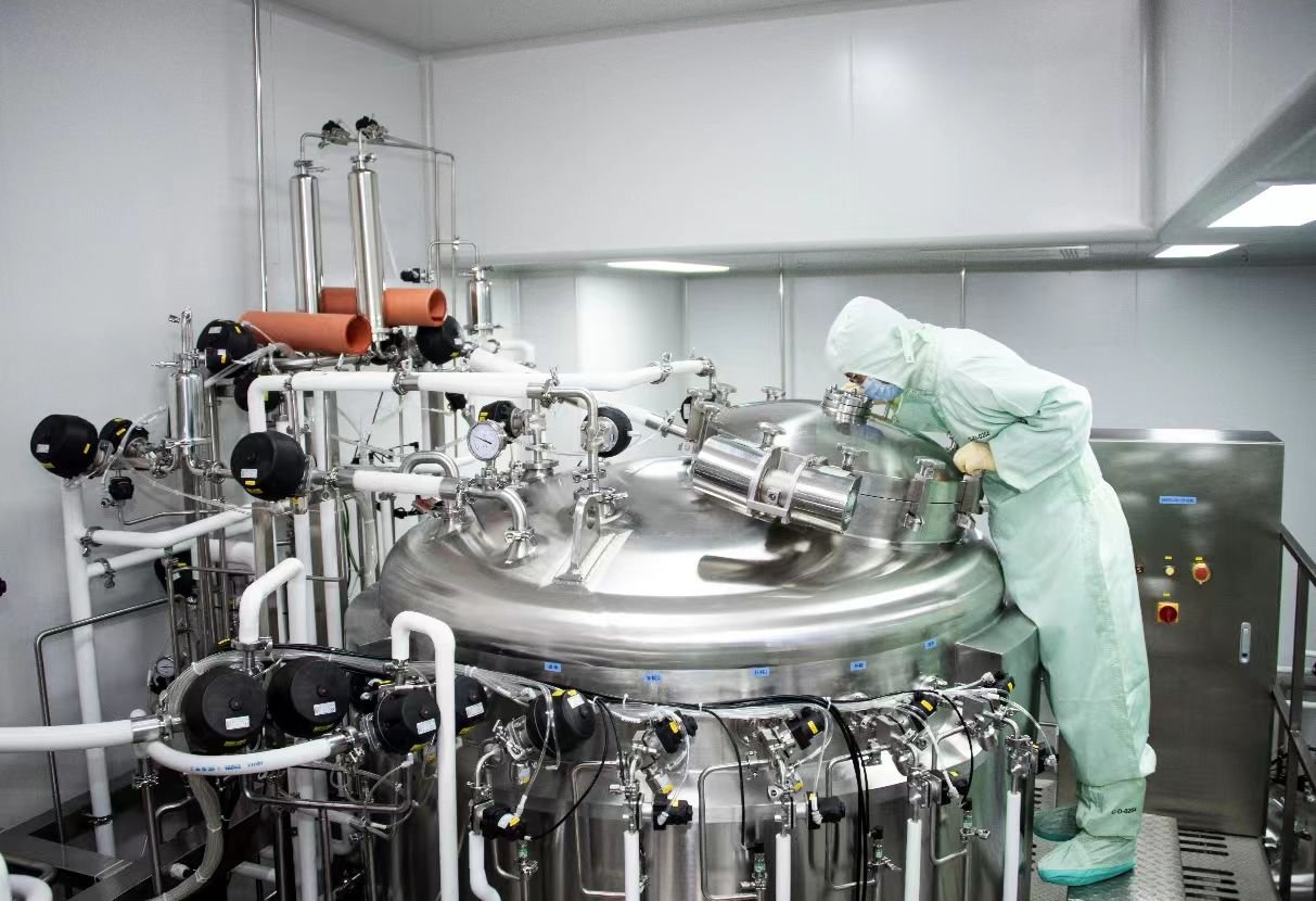
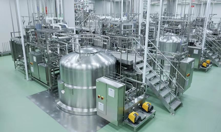
3.3 Large-Scale Bioreactor Cultivation
After multi-stage seed expansion, cells are transferred into production bioreactors for large-scale cultivation. All core process parameters including temperature, CO₂ level, DO, agitation rate and pH are continuously controlled throughout the culture period. Upon completion of cultivation, cell culture broth is subjected to centrifugation or depth filtration for initial separation prior to antibody purification.
3.4 Cell Culture Process Development Based on Quality by Design (QbD)
The core philosophy of Quality by Design (QbD) states that product quality is achieved through rational process design rather than relying solely on final testing. Since 2013, the U.S. Food and Drug Administration (FDA) has mandated the application of QbD principles for all new drug approval applications. Currently, QbD has become a consensus among domestic biopharmaceutical enterprises and is widely applied in the cell culture process development for monoclonal antibody products, in accordance with the guidelines outlined in ICH Q8.
The QbD-driven cell culture development workflow is as follows: First, define the Quality Target Product Profile (QTPP) as the starting point of R&D. Based on the understanding of Critical Material Attributes (CMAs), DOE is applied to identify Critical Quality Attributes (CQAs) and determine Critical Process Parameters (CPPs). A robust Design Space (DS) is established by evaluating the impacts of raw material properties, process conditions and environmental factors. Subsequently, quality risk management systems, control strategies and overall pharmaceutical quality management systems are implemented. This systematic approach deepens the understanding of products and manufacturing processes, enhances the stability of monoclonal antibody products, and reduces the complexity and costs of GMP-compliant production.
Antibody expression is a core procedure following cell cultivation, mainly consisting of transfection, clonal selection and cell amplification.
Transfection refers to the introduction of target antibody genes into host cells to enable the expression of recombinant proteins, which is divided into transient transfection and stable transfection. Prior to stable cell line screening, transient transfection is commonly used to generate preliminary antibody samples for quality assessment, as well as to optimize genetic elements such as promoters and codons.
In stable transfection, exogenous DNA can either integrate into the host chromosome or exist as episomal DNA. For CHO cell lines, episomal DNA will be lost during serial passaging, while chromosomally integrated exogenous genes can be stably inherited. Common transfection methods for mammalian cells include calcium phosphate transfection, cationic lipofection, cationic polymer transfection and electroporation, among which cationic lipofection and electroporation are the most widely used.
Screening high-yield single-cell clones is the key step in cell line development. Selection criteria include antibody titer, product quality, metabolic stability and cell line stability. After target genes are transfected into host cells and cultured under selective pressure, a mixed cell pool is obtained. The expression level of the cell pool is affected by transfection efficiency, selective pressure and the random integration pattern of exogenous genes. Significant variations in antibody titer are commonly observed among different cell populations, necessitating further screening to isolate high-producing and stable clones.
After CHO cell transfection, a bulk cell pool is first generated for selective screening. Selection strategies rely on the metabolic pathways of antibiotics or selective agents. The most frequently used screening reagents include puromycin, G418, methotrexate (MTX) and methionine sulfoximine (MSX):
Puromycin: An aminoglycoside antibiotic that inhibits protein synthesis by interfering with ribosomal function. The pac gene confers resistance to puromycin, and the working concentration ranges from 10 to 50 μg/mL.
G418: Another widely used aminoglycoside antibiotic for stable transfection screening, with a typical working concentration of 200 to 1000 μg/mL.
MTX: A folate antagonist that inhibits dihydrofolate reductase (DHFR) activity and nucleic acid synthesis. Gradual increase of MTX concentration induces DHFR gene amplification, which elevates the copy number of linked target genes and enhances antibody expression. The applicable concentration is 25 to 1000 nmol/L.
MSX: Used in the glutamine synthetase (GS) amplification system, an efficient expression amplification platform with superior amplification effects. The working concentration ranges from 25 to 500 μmol/L.
Major single-clone isolation techniques include Limiting Dilution Cloning (LDC), Fluorescence-Activated Cell Sorting (FACS) and semi-solid medium screening. Limiting dilution cloning is widely adopted due to its low cost and simple operation. Cells are diluted to an extremely low density and seeded into 96-well plates to ensure less than one cell per well. Microscopic imaging is performed to verify monoclonality. After incubation, high-titer clones are identified via ELISA and expanded. This procedure is usually repeated twice to guarantee clonal purity.
Fluorescence-activated cell sorting has gained increasing popularity. Cells secreting target antibodies are incubated with fluorescently labeled secondary antibodies, and surface-bound antibodies are detected and sorted by flow cytometry. Hydrogel microencapsulation is applied to retain secreted antibodies on the cell surface for improved screening efficiency. An advanced FACS strategy co-transfects host cells with two separate vectors encoding split GFP fragments fused to antibody heavy and light chain genes. Complete GFP fluorescence is only generated when both antibody chains are expressed, enabling high-throughput screening of high-yield clones as early as 48 hours post-transfection and substantially shortening the screening cycle.
Monoclonal antibodies, the most extensively applied therapeutic proteins, are predominantly manufactured via CHO cell culture. Compared with earlier biotech products such as cytokines, antibody drugs require large clinical dosages, which demands large-scale industrial production. Process optimization, scale-up and consistent high titer production remain major challenges for the domestic antibody industry. Improving cell culture titers, expanding production scale and maintaining stable product quality have become urgent priorities.
The primary objectives of antibody purification are to separate target antibodies from process-related and product-related impurities, and to obtain final products with high purity and low safety risks. Process-related impurities to be removed include intact cells, cell debris, host cell proteins (HCP), host cell DNA and residual medium components. Product-related impurities mainly consist of antibody fragments and aggregates. In addition, purification processes must possess robust viral clearance and inactivation capacity to eliminate endogenous virus-like particles and adventitious viruses. The core purification technologies include affinity chromatography, ion exchange chromatography and size-exclusion chromatography.
5.1 Cell Harvest, Centrifugation and Filtration
Antibodies are secreted extracellularly, so the first purification step is to separate cell culture supernatant from cells and cell debris. Bench-scale experiments commonly use batch centrifugation, while large-scale production adopts continuous separation technologies including depth filtration, tangential flow filtration (TFF) and continuous flow centrifugation.
Depth filtration features simple operation and low upfront investment. Filter media with broad pore size distribution remove particulate contaminants via both size exclusion and surface adsorption, enabling high throughput. The filter train is configured with media of gradually decreasing pore size from upstream to downstream. Since depth filtration cannot achieve sterile filtration, a final sterile filter is integrated into the system. Diatomaceous earth contained in depth filter media carries positive charges, which can partially adsorb negatively charged host cell DNA and HCP for preliminary purification. However, disposable depth filter cartridges incur high operational costs and have limited loading capacity. Parallel installation of multiple filters is required for large-volume processing, increasing footprint and expenses. This technology is mainly applied to pilot-scale purification with working volumes ranging from 100 L to 2000 L.
Tangential flow filtration is widely used in perfusion bioreactor systems. High cross-flow velocity minimizes cell deposition on membrane surfaces and prevents clogging during large-volume processing. Nevertheless, excessive cross-flow velocity generates high shear force, which may rupture cells and release intracellular proteins and nucleic acids, increasing the burden on downstream purification. Hollow fiber membranes are the dominant format for TFF, featuring large surface area and controllable shear force. Process scale-up is realized by increasing the number of hollow fibers. Key parameters for TFF optimization include membrane material, surface area, pore size, cross-flow rate and transmembrane pressure. A small volume of buffer can be added at the final stage of filtration to reduce dead volume and minimize antibody loss. TFF supports ultra-large-scale processing (over 10,000 L) with a throughput up to nearly 5000 L per hour. Its limitations include high membrane costs, long processing time and limited flow rate controllability.
Continuous flow centrifugation, especially disc stack centrifuges, is the mainstream solid-liquid separation technology for commercial-scale antibody production. Cell culture broth is fed into the centrifuge chamber, and cells are sedimented along the conical wall under centrifugal force. Accumulated cells are periodically discharged via brief valve opening, and the liquid loss during cell harvesting can be controlled below 5% by optimizing rotational speed, discharge frequency and duration. Disc stack centrifuges offer advantages including compact footprint, large sedimentation area, high throughput, reliable operation and low long-term operational costs. The main drawbacks are high upfront equipment investment, complex cleaning procedures and the lack of lab-scale mimicking models. This technology achieves a cell removal efficiency of over 90% and a perfusion rate up to 3600 L/d. Attention must be paid to cell tolerance to shear force to avoid cell damage during operation. Residual fine cell debris after centrifugation can be further removed by depth filtration.
5.2 Affinity Chromatography
Affinity chromatography separates target molecules based on specific biological interactions between analytes and immobilized ligands. Protein A and Protein G are the most commonly used affinity ligands for antibody purification. The separation principle relies on specific biomolecular interactions including antigen-antibody binding. Target proteins are captured by ligands immobilized on chromatography media via covalent bonds, van der Waals forces, hydrophobic interactions and electrostatic forces. This technology delivers high purification efficiency and excellent specificity.
Native Protein A, a surface protein derived from Staphylococcus aureus, contains five IgG-binding domains and additional non-Fc-binding regions, with a molecular weight of 42 kDa. It exhibits strong binding affinity toward the Fc region of IgG molecules. However, non-specific binding caused by extra domains reduces product purity. Genetically engineered recombinant Protein A with truncated non-essential domains is therefore widely used in commercial chromatography resins to improve purification performance. Resins with truncated B domains enable high-purity IgG recovery under mild elution conditions, preventing protein aggregation and preserving biological activity.
Over two-thirds of commercially approved antibody drugs adopt Protein A affinity chromatography as the primary capture step. Protein A specifically binds to the Fc region of monoclonal antibodies and removes the majority of impurities including HCP, host DNA and potential viruses in a single step, achieving a product purity above 95% and nearly 100% recovery yield. Cell culture supernatant can be loaded directly onto Protein A columns without pre-treatment. The standard workflow includes column loading, washing to eliminate weakly bound impurities, elution using acidic buffer (pH 3.0–3.5), stringent cleaning with stronger acid (pH ~2.0) to remove tightly bound contaminants, and column regeneration. Common cleaning reagents include guanidine hydrochloride, urea and dilute alkaline solutions. Protein A chromatography also serves as an efficient concentration step, with eluate antibody concentrations exceeding 10 g/L and reducing the processing volume for subsequent polishing steps.
Key parameters for Protein A process optimization include dynamic binding capacity, residence time (flow rate), washing and elution conditions. Significant variations are observed among different monoclonal antibodies: dynamic binding capacity ranges from 10 to 40 g/L (a 3-fold difference), and elution pH varies from 3.0 to 4.1. Residual HCP levels and aggregate content in eluates also differ substantially, indicating that customized process optimization is required for individual antibody products even on universal platform workflows.
Proteolytic enzymes in cell culture supernatant cause Protein A leakage during loading. Since dissociated Protein A possesses immunogenicity, additional polishing steps are necessary for its removal. Despite its superior performance, Protein A has notable disadvantages: extremely high resin cost (approximately 10 times higher than conventional chromatography media), limited dynamic binding capacity (20–30 g/L), restricted linear flow rate and the requirement for low-pH elution. To control production costs, Protein A resins are reused for up to 300 cycles with mild cleaning protocols. Large-scale manufacturers often use relatively small Protein A columns and process harvest broth in multiple batches, which limits overall purification throughput. A dual-flow-rate loading strategy is proposed to improve efficiency: high flow rate at the initial loading stage for accessible binding sites, followed by reduced flow rate to allow sufficient diffusion for full resin utilization.
Native Protein A is susceptible to degradation under strong alkaline conditions and can only be cleaned with dilute NaOH (~0.1 mol/L), which has limited endotoxin removal capacity. MabSelect SuRe (GE Healthcare), an engineered Protein A variant with modified alkali-labile asparagine residues, exhibits excellent tolerance to strong alkaline cleaning. Meanwhile, research is ongoing to develop alternative ligands including peptides, aptamers and synthetic molecules. Nevertheless, these alternatives cannot yet match Protein A in binding affinity, specificity and versatility, and Protein A will remain the dominant capture ligand for antibody purification in the next 5 to 10 years.
5.3 Ion Exchange Chromatography
Ion exchange chromatography is the most widely applied polishing technology, which separates molecules based on differences in surface charge by adjusting buffer pH and ionic strength. It should be noted that chromatographic interactions depend on local surface charge rather than the overall isoelectric point (pI) of proteins. Cation exchange media bind positively charged molecules, while anion exchange media retain negatively charged molecules.
Cation exchange chromatography has been adopted as the primary capture step for multiple commercial therapeutic antibodies including Humira, Synagis, Soliris and Zenapax, as well as numerous clinical-stage candidates. Compared with Protein A, cation exchange resins deliver distinct cost advantages: the resin price is merely one-tenth of Protein A media, and the resins can be reused over 100 times with strong alkali resistance. Their dynamic binding capacity reaches up to 100 g/L, 2 to 5 times that of Protein A, lowering resin consumption. A two-step purification workflow combining cation exchange capture and anion exchange polishing achieves an overall recovery of 82% and product purity above 97%, and is scalable up to 1000-fold.
The main limitations of cation exchange chromatography are lower specificity and versatility. Process conditions must be individually optimized according to the pI of each antibody, and post-translational modifications also lead to variations in surface charge, affecting buffer pH selection.
After capture, additional polishing steps are required to further remove HCP, host DNA, antibody aggregates, fragments and leached Protein A to meet safety and purity specifications. Most manufacturing processes adopt two polishing steps for sufficient purification redundancy, while single-step polishing workflows are increasingly developed to improve efficiency. Common polishing techniques include anion exchange chromatography, cation exchange chromatography, hydrophobic interaction chromatography (HIC) and hydroxyapatite chromatography. Anion exchange and HIC are typically operated in flow-through mode, whereas cation exchange and hydroxyapatite chromatography use bind-elute mode. Polishing resins with small particle sizes (10–30 μm) are applied to enhance resolution, at the cost of reduced flow rate and prolonged processing time.
The classical polishing workflow combines flow-through anion exchange and bind-elute cation exchange: anion exchange efficiently removes host DNA and HCP, while cation exchange is effective for eliminating aggregates and antibody fragments. The net charge of antibodies is determined by solution pH relative to their pI: antibodies carry positive charges when buffer pH is below the pI, and negative charges when pH is above the pI. Strong ion exchange media are predominantly used in antibody purification due to their pH-independent charge properties.
The standard operating procedure for ion exchange chromatography includes column equilibration, sample loading, washing, elution and regeneration. Low-ionic-strength buffers are used for loading to promote analyte binding. Elution is achieved by increasing salt concentration to enhance competitive ion displacement. Linear gradient elution improves resolution, while stepwise elution is preferred in large-scale production for operational stability.
Weak Partition Chromatography (WPC), a modified anion exchange operation between pure flow-through and bind-elute modes, has been developed for single-step polishing. Low ionic strength enhances impurity binding while causing partial antibody retention, which can be recovered by brief washing. A combined workflow of Protein A capture and WPC polishing can meet the clearance requirements for HCP, leached Protein A, endotoxin and host DNA. Aggregate removal is case-dependent: aggregates are retained on anion exchange media for certain antibodies, while monomeric antibodies bind preferentially for others, requiring customized resin selection and operating modes.
Membrane chromatography, especially anion exchange membranes, has seen rapid adoption in recent years. With identical functional groups as traditional resin media, membrane chromatography features convection-driven mass transfer, enabling ultra-high flow rate and dynamic binding capacity (1–10 kg/L membrane volume, 10 to 100 times higher than resins). It also reduces buffer consumption and equipment footprint, with a recovery yield of 95%–100% and equivalent performance in removing HCP, DNA and viruses. The main drawback is higher costs for single-use membrane devices compared with reusable resins.
5.4 Size-Exclusion Chromatography (SEC)
Also known as gel filtration chromatography, SEC separates molecules based on molecular size. Porous gel matrices exclude large molecules, which migrate rapidly through interstitial spaces, while small molecules penetrate into gel pores and exhibit longer retention time. Dextran gels (Sephadex) are the most commonly used media. Major applications include fractionation of antigens and antibodies, removal of small molecular impurities such as salts and peptide fragments, and molecular weight determination.
After primary purification, final polishing is performed using advanced technologies such as countercurrent chromatography and High-Performance Liquid Chromatography (HPLC). Various HPLC modes are applied for the characterization and fine purification of antibody products: Size-Exclusion HPLC (SE-HPLC) analyzes aggregates and fragments; Ion-Exchange HPLC (IE-HPLC), Hydrophobic Interaction HPLC (HI-HIC) and Reversed-Phase HPLC (RP-HPLC) resolve charge variants, structural isoforms and heterogeneous species induced by glycosylation, deamidation and oxidation.
6. Antibody Drug Formulation
The final manufacturing stage is the formulation of purified antibodies into finished drug products, covering formula design, stability evaluation and sterilization.
Formulations are rationally designed according to the biochemical characteristics of target antibodies. A typical antibody formulation consists of the active pharmaceutical ingredient (antibody), buffering agents, tonicity modifiers, lyoprotectants and surfactants. Common tonicity modifiers include sodium chloride, disaccharides (sucrose, trehalose, maltose) and sugar alcohols (mannitol, sorbitol). Sucrose and trehalose are the primary lyoprotectants for freeze-dried products. The pH range of liquid antibody formulations is controlled between 4.0 and 8.0, with buffering systems including histidine, citrate, succinate, acetate, phosphate, glutamate and TRIS. Polysorbate 20 and Polysorbate 80 are the most frequently used surfactants.
6.2 Antibody Stability Study
Comprehensive stability testing is conducted to evaluate product performance under various storage conditions including temperature and light exposure. Poor antibody stability leads to reduced in vivo efficacy in clinical trials. Structural stability is the foundation for antibody affinity and specificity. Stable protein conformation also minimizes misfolding during biosynthesis and increases soluble expression levels. Improved thermal stability reduces the exposure of protease cleavage sites, extends shelf life and optimizes storage conditions.
Major strategies to enhance protein stability include non-covalent modification, chemical modification, stabilizer supplementation, protein engineering and biomineralization. Multiple intrinsic and extrinsic factors affect antibody stability:
(1) Intrinsic molecular properties
Amino acid sequences determine the aggregation propensity of antibodies. Extreme pI values of the complementarity-determining region (CDR) promote aggregation: antibodies with low pI form soluble aggregates via intermolecular electrostatic interactions, while high-pI antibodies tend to form precipitates when contacting hydrophobic surfaces. Distinct IgG subclasses also exhibit different stabilities under acidic stress: IgG1 maintains monomeric state, whereas IgG2 and IgG4 are prone to dimerization and irreversible aggregation.
(2) Environmental stress factors
Temperature: Elevated temperature irreversibly disrupts antibody conformation and accelerates deamidation and oxidation. The melting temperature (Tm) of most antibodies ranges from 40 °C to 80 °C, while standard storage conditions (2–8 °C) are well below this threshold. Repeated freeze-thaw cycles also cause protein denaturation due to pH fluctuation, solute concentration and ice-water interface effects.
Light exposure: Aromatic amino acid residues (primarily tryptophan) are susceptible to photo-oxidation, leading to protein cleavage and cross-linking. UV light causes more severe damage than visible light. Lyophilized formulations demonstrate better photostability than liquid formulations. Photodegradation of the CH2 domain is observed even under standard ICH Q1B light exposure conditions.
Mechanical stress: Agitation, pumping and filtration generate shear force and increase air-liquid interface area, inducing protein unfolding and aggregate formation (particle size: 1.5–80 μm). High-concentration antibody solutions exhibit shear thinning behavior.
(3) Formulation and packaging factors
High antibody concentration enhances intermolecular interactions and aggregation. Surfactants such as Polysorbate 20 reduce protein adsorption to packaging materials. Polyvinyl chloride (PVC) containers cause more severe antibody adsorption and particle formation than polyolefin materials.
Mechanisms of protein instability
Instability is categorized into chemical instability and physical instability, which interact with each other:
Chemical degradation: Oxidation of methionine, histidine and cysteine residues; deamidation of asparagine and glutamine; hydrolysis of peptide bonds and disulfide bonds; Maillard reaction induced by reducing sugars. Degradation occurring in the CDR region significantly impairs antibody affinity, while modifications in the Fc region accelerate in vivo clearance.
Physical instability: Protein denaturation and aggregation. Aggregates are formed via non-covalent interactions or covalent disulfide cross-linking. Large antibody aggregates possess strong immunogenicity, potentially triggering adverse immune responses and hypersensitivity reactions in patients.
6.3 Stability Assessment Technologies
Stability evaluation combines biological potency assays, structural and purity analysis, and routine quality testing (appearance, pH, etc.). Common analytical techniques include Enzyme-Linked Immunosorbent Assay (ELISA), Differential Scanning Calorimetry (DSC), Differential Scanning Fluorimetry (DSF), Circular Dichroism (CD) spectroscopy and Dynamic Light Scattering (DLS). High-throughput technologies such as Cross-Interaction Chromatography (CIC), AC-SINS and CSI-BLI predict self-association propensity of antibodies. In addition, computer-aided molecular modeling and molecular dynamics (MD) simulations are applied to analyze antibody structure, binding interactions and energy distribution.
The entire manufacturing workflow of antibody drugs covers cell cultivation, antibody expression, purification and formulation. Each process step is indispensable for production. Strict process control and systematic optimization across all stages ensure the manufacture of high-purity, high-quality antibody drugs for clinical disease treatment.
As a leading process-oriented engineering company, Sino Bioengineering delivers professional bioengineering solutions and comprehensive technical services to clients worldwide.
For more products and solutions: https://sinobioengg.com/category/solution/
For more service: https://sinobioengg.com/category/service/