

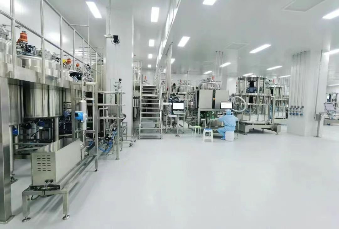
With successive breakthroughs of nucleic acid therapeutics in the treatment of diverse diseases and the clinical translation of mRNA vaccines and small interfering RNA therapeutics, nucleic acid formulations have emerged as a “golden track” within biopharmaceuticals. Although numerous candidate vaccines for infectious disease prevention and cancer therapy have entered clinical trials, their clinical translation remains hindered by insufficient targeting accuracy, inherent immunogenicity, toxicity risks, and limitations of universal delivery platforms. This article focuses on delivery optimization strategies to address existing bottlenecks and outlines future directions to drive relevant advances.
Unprotected mRNA undergoes rapid degradation by nucleases in vivo, resulting in a short half-life. Furthermore, negatively charged mRNA generates electrostatic repulsion with anionic cell membranes, impeding cytoplasmic internalization. In addition, unmodified exogenous mRNA can be recognized by pattern recognition receptors (PRRs), triggering robust innate immune responses. Efficient delivery systems are therefore indispensable to tackle these challenges. An ideal carrier should possess the core capabilities to encapsulate and shield mRNA, enhance cellular uptake, facilitate effective endosomal escape, and release mRNA efficiently into the cytoplasm for translation. Multiple mRNA delivery platforms have been engineered to date, including polymeric nanoparticles and liposomes. This article elaborates delivery optimization strategies from two perspectives:
1.Optimizing LNP formulations (component ratio tuning, SORT strategy, etc.) to modulate immune responses and achieve organ-specific targeting;
2.Surface functionalization of LNPs (conjugation with small molecules, peptides and antibodies) to enable precise targeting of specific cells or organs.
Composition of LNPs
Lipid nanoparticles (LNPs) typically range from 100 to 200 nm in diameter, consisting of ionizable lipids, helper phospholipids, cholesterol, and PEG-lipids (Figure 1). Ionizable cationic lipids account for approximately 50 mol% of LNP formulations. Multiple structural scaffolds are designed for the head groups of ionizable lipids, including tertiary amines, branched polyamines and cyclic moieties (e.g., piperidine, diketopiperazine or benzene rings), which govern the acid dissociation constant (pKₐ) of ionizable lipids. The pKₐ value determines LNP surface charge, stability and delivery efficiency. Critically, pKₐ serves as a key regulator of organ tropism: hepatic-targeted LNPs exhibit a pKₐ of 6–7, lung-targeted LNPs have a pKₐ above 9, while spleen-targeted LNPs feature relatively low pKₐ values.
Helper phospholipids constitute 10–20 mol% of total lipids. They facilitate the formation of stable lipid bilayers and improve particle stability via elevated phase transition temperatures. Widely adopted phospholipids such as DSPC and DOPE play vital roles in regulating delivery performance. For instance, studies have demonstrated that DOPE improves mRNA transfection efficiency. Moreover, phospholipids with distinct chemical properties alter the biodistribution of LNPs. Neutral phospholipids tend to accumulate in the liver, whereas anionic phospholipids confer superior spleen-targeting capacity; incorporation of bioactive phospholipids can further enhance organ selectivity.
Cholesterol generally makes up 38 mol% of lipid components. As an essential constituent of cell membranes, cholesterol’s stereochemical structure and tailored modifications exert profound regulatory effects on the stability, fluidity and permeability of LNPs.
PEG-lipids account for less than 2 mol%. PEGylated lipids are formed by conjugating hydrophobic alkyl chains with hydrophilic polyethylene glycol (PEG), establishing a steric barrier on the LNP surface that promotes accumulation at target sites, prevents particle aggregation and maintains optimal particle size (typically 50–120 nm; formulations lacking PEG are unstable and readily exceed 200 nm). Nevertheless, excessive PEG hinders endosomal fusion and restricts cellular uptake and mRNA release. Additionally, a prominent drawback associated with widespread PEG utilization is the high prevalence of anti-PEG antibodies. Blood samples collected in 2019 revealed positive rates as high as 83% among donors.
LNP Preparation and Characterization
Particle size (commonly 20–200 nm) is a decisive parameter governing the delivery performance of mRNA-LNPs, including cellular uptake, biodistribution, immunogenicity and clearance kinetics. Precise size control is essential for targeted delivery to specific tissues and cells, improved tissue penetration and prolonged in vivo retention. mRNA-LNPs are predominantly manufactured via microfluidic mixing or ethanol dilution methods. Both approaches rely on the self-assembly of lipids (dissolved in ethanol) and mRNA (in acidic buffer at pH ≈ 4.0), followed by ethanol removal, pH neutralization (to pH 7.4) and sterile filtration. Mixing conditions exert a dominant influence on particle characteristics. Despite straightforward operation, the ethanol dilution method often generates broad particle size distributions and moderate encapsulation efficiency (70–85%). By contrast, microfluidic mixing enables rapid, homogeneous blending of lipid-ethanol solutions and aqueous mRNA phases. Research also indicates that optimal encapsulation and cellular expression are achieved when mRNA solution pH is maintained at 4.0; supplementing storage buffer with sucrose (optimal concentration: 300 mM) mitigates aggregation and mRNA leakage to improve freeze-thaw stability.
Comprehensive characterization of LNP parameters is mandatory to guarantee quality and efficacy, including particle size, polydispersity index (PDI), zeta potential and RNA encapsulation efficiency (EE). Dynamic light scattering (DLS) is applied to characterize LNP particle size, zeta potential and PDI. Acceptable LNPs generally display diameters of 50–200 nm with PDI < 0.3; lower PDI values correspond to more homogeneous particle populations. For mRNA-loaded LNPs, near-neutral surface charge is preferred. RNA EE (%) is quantified via the RiboGreen assay: the concentration of externally unencapsulated RNA is first measured, followed by lysis of LNPs using surfactants to determine the total RNA concentration (both encapsulated and free).
Optimization Strategies for mRNA-LNP Delivery Systems
1. LNP Formulation Optimization
As outlined in the preceding section, structural modification of the four core LNP components alters the overall stability, mRNA encapsulation efficiency, transfection efficacy and cytotoxicity of mRNA-LNPs. Studies have proven that tuning the ratio between phospholipids and PEG-lipids enables immune response modulation. LNPs formulated with DSPC and 0.5 mol% PEG-lipids elicit the strongest humoral immune responses, driving elevated production of binding antibodies, enhanced virus neutralization capacity, and robust responses of memory B cells (MBCs) and long-lived plasma cells. LNPs incorporating DOPS and 1.5 mol% PEG-lipids deliver optimal cellular immunity, markedly boosting the generation of SARS-CoV-2-specific CD8⁺ T cells secreting interferon-γ (IFN-γ) and tumor necrosis factor-α (TNF-α).
Growing research efforts focus on introducing a fifth component into conventional four-component LNPs. The group led by Daniel J. Siegwart incorporated DOTAP, a fifth molecule with defined charge properties, to adjust LNP surface charge for precise organ targeting: formulations containing 0% DOTAP favor hepatic delivery; 10–15% DOTAP facilitates spleen targeting, while a 50% DOTAP ratio achieves optimal lung delivery.
In contrast to additive strategies, simplified LNP formulations have also demonstrated improved targeting performance. Researchers discovered that cholesterol and phospholipids are not strictly required for LNP functionality. Three-component LNPs (ionizable cationic lipid, permanent cationic lipid and PEG-lipid) can accomplish specific lung targeting.
2. Surface Modification of Lipid Nanoparticles
Active targeting can be realized through chemical modification of LNPs, representing a critical strategy to enhance therapeutic precision and efficacy. Active targeting is generally implemented via surface functionalization of LNPs with diverse ligands capable of selective binding to receptors overexpressed on target cells. This approach elevates mRNA concentrations in target organs and minimizes off-target adverse effects. Ligands fall into two major categories based on molecular weight: small molecules (carbohydrates, folate, mannose) and macromolecules (antibodies, peptides, aptamers).
Folic acid (FA) is a widely investigated small-molecule targeting ligand in clinical research, exhibiting high affinity for folate receptors frequently upregulated in multiple human malignancies. Folate-conjugated nanoparticles or liposomes undergo active internalization via folate receptor-mediated endocytosis, enabling effective targeting of folate receptor-positive tumor cells. Certain human cancers, including prostate cancer, overexpress σ receptors, and benzamide analogues display high affinity toward these receptors. This strategy has been broadly adopted in drug delivery. Beyond direct tumor cell targeting, immune cells such as dendritic cells (DCs) constitute pivotal targets for cancer therapeutics.
Owing to exceptional specificity and diversity, antibodies serve as ideal targeting ligands for LNP modification to achieve precise mRNA delivery. Antibodies can be conjugated to LNPs via Fc-binding peptides, surface linkers or covalent chemical bonds. Appropriate antibody selection depends on antigens highly expressed on target cell surfaces. Antibody-modified LNPs represent the prevailing strategy for targeting non-hepatic organs. Anti-HER2 antibody-conjugated LNPs developed by BioNTech effectively target breast cancer lesions; BNT111 (anti-HER2-LNP mRNA vaccine) has advanced to Phase I/II clinical trials (NCT04503278). In vivo mRNA delivery systems targeting CD4⁺ T cells support rapid and robust generation of CAR-T cells, opening new avenues for in vivo CAR-T therapy.
Conjugating targeting antibodies onto LNP surfaces via thiol-maleimide reaction constitutes the mainstream workflow for antibody-LNP (Ab-LNP) preparation. Tris(2-carboxyethyl)phosphine hydrochloride (TCEP) is a low-toxicity, high-efficiency thiol reductant widely deployed in biopharmaceuticals to stabilize proteins and nucleic acids. Antibody proteins are incubated with TCEP at ambient temperature at a defined molar ratio to reduce disulfide bonds. The activated antibodies are subsequently reacted with LNPs bearing maleimide groups (where a fraction of DMG-PEG is substituted with Mal-PEG2K-DSPE) under room temperature, yielding Ab-LNPs under mild and efficient reaction conditions.
Conclusion
Process development for LNP delivery systems essentially relies on the synergy between precision control and functional prioritization: formulation and process tuning are required to construct stable, homogeneous delivery carriers, while the core capability of efficient nucleic acid delivery must be maintained with clinical demands as the primary focus. Future breakthroughs will rely on multidisciplinary innovation, concentrating on the development of degradable lipids and novel targeting ligands to boost delivery precision; adoption of more biocompatible polymers (such as pSar and POx) to replace PEG for improved safety; and utilization of artificial intelligence (AI) to accelerate LNP formulation design and performance prediction. With continuous technological iteration, LNPs will evolve beyond mere delivery vehicles for nucleic acid therapeutics and become the core engine enabling precision treatment. They will accelerate the translation of numerous nucleic acid therapeutics from laboratory benches to clinical practice and bring transformative advances for disease management.