Particle size stands as a make-or-break critical parameter in the in vivo delivery journey of mRNA-LNP therapeutics. Unlike mRNA, which encodes biological instructions, or ionizable lipids engineered for pH-responsive performance, even minor nanometer-scale shifts in particle diameter exert profound impacts on every performance metric of LNPs, including colloidal stability, mRNA encapsulation efficiency, in vivo biodistribution, and cellular uptake. What constitutes the ideal particle size for LNPs, and what scientific rationale underpins this critical numerical range?
The Consensus Gold Standard Window: 60–100 nm
Data from all commercialized mRNA vaccines and extensive academic research converge on a narrow particle size range of 60 to 100 nm for LNPs. The two leading SARS-CoV-2 mRNA vaccines both exhibit mean particle diameters between 80 and 100 nm, with the formulations developed by Pfizer/BioNTech and Moderna falling squarely within this spectrum. Manufactured via microfluidic mixing technology, lipid-mRNA complexes routinely yield particles sized 60–100 nm. For instance, the SARS-CoV-2 vaccine candidates nCoVsaRNA and ARCoV display average particle sizes of 75 nm and 89 nm, respectively.
This range is not arbitrarily selected but represents an optimal balance constrained by interrelated physical, chemical, and biological factors. The U.S. FDA classifies particle size and particle size distribution as Critical Quality Attributes (CQAs) for lipid nanoparticle pharmaceutical products. The industry-wide consensus specifies an ideal particle size of 60–100 nm paired with a polydispersity index (PDI) below 0.2. A systematic review further confirms that established LNP manufacturing platforms can stably generate particles spanning 60–180 nm with customizable diameters, while a PDI ≤ 0.30 serves as the baseline regulatory compliance threshold for pharmaceutical-grade formulations. Collectively, the 60–100 nm window has become universally recognized as the gold standard particle size for mRNA-LNP formulation development.
Drawbacks of Suboptimal Small Sizes: Renal Clearance and Reduced Encapsulation Efficiency
LNPs with diameters below 60 nm introduce two major liabilities. First, smaller particles possess limited internal volume to encapsulate mRNA payloads. Equivalent lipid input yields a greater number of miniature particles required to carry sufficient therapeutic mRNA, inflating formulation volume and manufacturing costs. Second, particles smaller than 50 nm become susceptible to rapid glomerular filtration. While the renal glomerulus primarily clears molecules under 10 nm, particles below 50 nm experience accelerated systemic renal elimination, shortening blood circulation half-life and compromising tissue-targeting capacity.
More critically, reduced particle size correlates with diminished encapsulation efficiency. Miniaturized LNPs lack sufficient internal lipid compartmentalization to sequester and shield mRNA cargo, leaving a substantial fraction of RNA exposed to extracellular nucleases and prone to degradation.
Conversely, particles exceeding 200 nm trigger distinct limitations: they are readily recognized and rapidly phagocytosed by macrophages resident in the liver and spleen, hindering delivery to target tissues. Professional phagocytes exhibit high sensitivity toward particulate foreign bodies, sequestering oversized LNPs within reticuloendothelial system (RES) organs rather than enabling specific tissue targeting. Undersized particles suffer inadequate cargo protection and rapid metabolic clearance, while oversized particles undergo premature phagocytic elimination—rendering 60–100 nm the balanced sweet spot between these two biological barriers.
Physiological Mechanisms Governing Size-Dependent Trafficking: Hepatic Sinus Fenestrae and Cellular Endocytosis
The 0–100 nm particle size range aligns precisely with anatomical microstructures governing hepatic and cellular access.
Following intravenous administration, most LNPs must traverse hepatic sinusoidal endothelium to reach hepatocytes. Hepatic sinus walls feature fenestrae with diameters ranging from 100 to 200 nm. LNPs larger than 200 nm cannot permeate these fenestral pores, whereas particles maintained below 100 nm readily extravasate into the hepatic parenchymal space. This physiological constraint explains why LNP-siRNA therapeutics (e.g., Onpattro) and most liver-targeted mRNA modalities lock particle size below 100 nm.
At the cellular level, particle diameter directly modulates endocytic uptake efficiency, the primary route by which therapeutic LNPs enter target cells. Endocytosis exhibits stringent size selectivity, with an optimal window of 80–120 nm for maximal internalization. Notably, influenza virions (~80 nm) and reoviruses (~120 nm) naturally fall within this span, an evolutionary adaptation enabling efficient receptor-mediated cellular invasion. LNPs sized 80–100 nm exploit this biologically conserved endocytic machinery honed by viral evolution to achieve robust cellular uptake.
Precision Tuning of Particle Size: Formulation and Manufacturing Strategies
LNP particle size is not a fixed product attribute post-fabrication and can be precisely modulated via multiple orthogonal approaches.
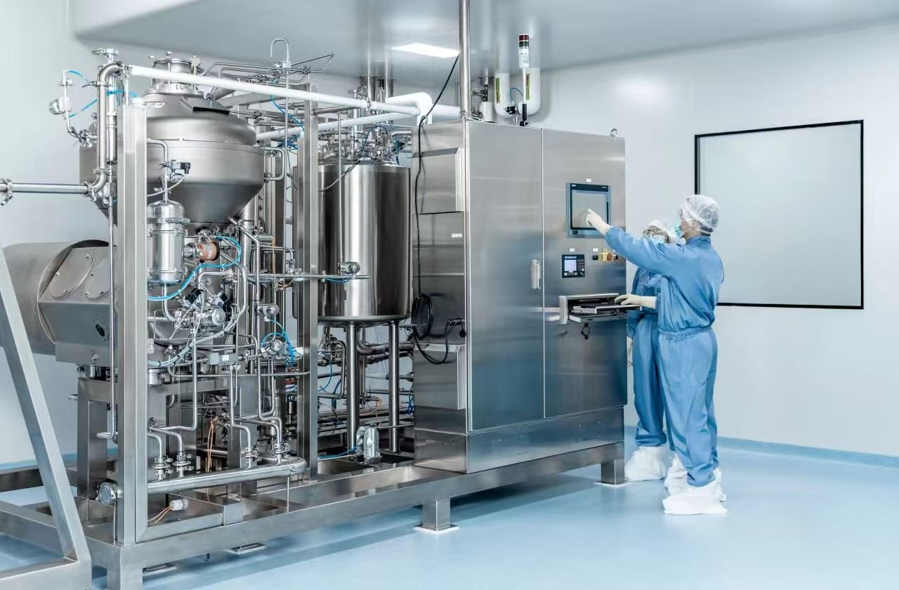
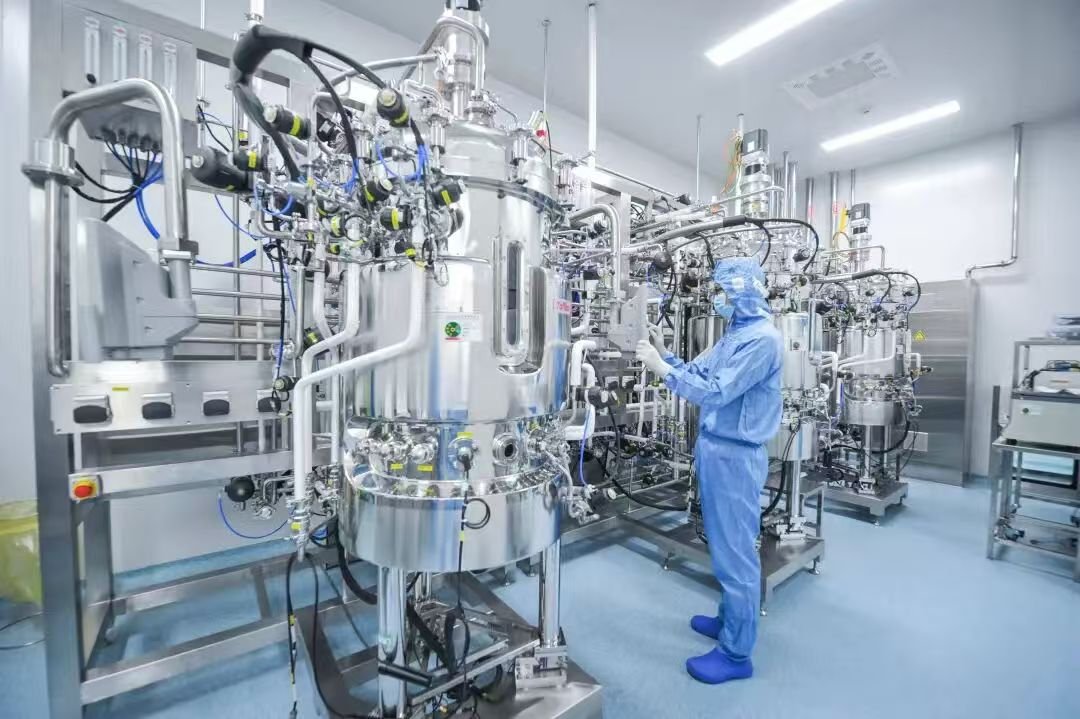
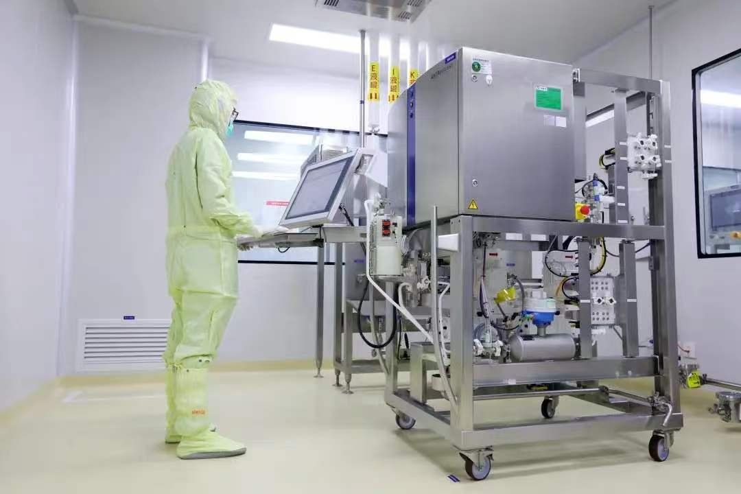
Microfluidic mixing remains the dominant industrial production platform for LNPs. Millisecond-scale mixing dynamics are finely tuned by adjusting flow rate ratios between the organic lipid-ethanol phase and aqueous mRNA acidic buffer phase, alongside total volumetric flow rates. Elevated flow rates accelerate rapid mixing to generate smaller particles, whereas reduced flow rates yield larger nanoparticles. Design of Experiments (DoE) methodologies further optimize process parameters to enable reproducible fabrication of custom-sized LNPs across the 60–180 nm spectrum.
Lipid formulation composition constitutes another core lever for size modulation. PEGylated lipids are typically incorporated at 1–2 mol%: excessive PEG-lipid elevates aqueous interfacial area and impairs encapsulation efficiency, while insufficient PEG-lipid promotes particle aggregation and size expansion. Fine adjustments to PEG molecular weight and the nitrogen-to-phosphate (N/P) ratio between ionizable lipids and mRNA also deliver granular control over particle diameter.
Novel size-modulation techniques have emerged in the early 2020s. Research demonstrates that buffer supplementation with monovalent salts (e.g., sodium chloride) offers a straightforward tuning strategy: salt addition enlarges particle diameters to above 200 nm in a concentration-dependent manner following Hofmeister series principles. Though LNPs sized 200–300 nm are suboptimal for intravenous systemic delivery, they facilitate robust dendritic cell uptake and have demonstrated promising performance in cancer vaccine applications.
Particle Size as a Determinant of Tissue Biodistribution
Differentially sized LNPs exhibit innate tissue tropism following systemic administration, a phenomenon termed passive targeting.
1.Particles <50 nm: Gradual accumulation within bone marrow
2.Particles 50–100 nm: Favorable hepatic tissue targeting
3.Particles 100–200 nm: Potent immunostimulatory capacity, ideal for vaccine platforms to activate antigen-presenting cells
4.Particles >200 nm: Mechanical entrapment within pulmonary capillaries, driving lung enrichment
This establishes particle diameter as a critical physical determinant dictating in vivo tissue distribution upon systemic injection.
Particle size transcends a mere numerical specification; it serves as the pivotal physical bridge connecting molecular lipid design to the in vivo fate of mRNA cargo. Spanning glomerular filtration thresholds, hepatic sinus fenestral dimensions, the optimal endocytic uptake window, and macrophage phagocytic cutoff limits, the 60–100 nm gold standard represents the most physiologically balanced range to overcome successive biological barriers. Elucidating the multifaceted impacts of particle size on delivery performance not only streamlines optimization of existing LNP formulations but also establishes foundational design principles for next-generation organ- and cell-specific targeted mRNA therapeutics. When LNPs navigate biological compartments at their physiologically optimized diameter, half the therapeutic success of mRNA medicines is already secured.