

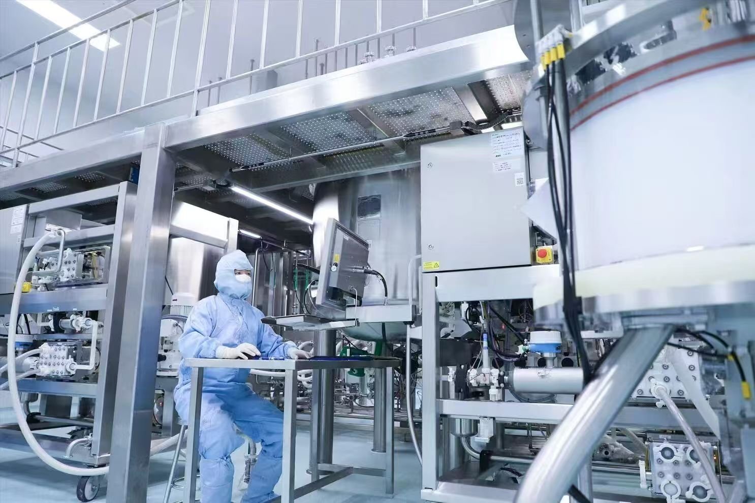
During downstream purification of antibody therapeutics, chromatographic steps – particularly affinity capture and polishing – directly determine product purity and yield. However, purification practitioners frequently encounter a troublesome issue: abnormal discoloration of chromatography columns, eluates or even final samples, ranging from pale yellow, tan and pink to dark green. Such discoloration not only compromises product appearance but also signals potential underlying process risks.
Why Pigment-Related Discoloration Requires Close Attention
1.Abnormal coloration serves as an early warning indicator of process variations. Fluctuations in medium batches, feeding strategies or cellular metabolic status can manifest as discoloration during chromatography.
2.Certain pigments (e.g., metal-phenolic complexes) may continuously leach in subsequent unit operations if not removed, triggering off-color specifications in final drug formulations and jeopardizing product quality.
3.Pigments often co-adsorb with host cell proteins (HCPs) and DNA. Elimination of pigments frequently enables concurrent clearance of these critical impurities.
Pigment remediation is challenging due to diverse origins and complex mechanisms. Some pigments originate from low-molecular-weight medium components (e.g., vitamins, metal ions), while others co-adsorb with HCP aggregates. Without targeted mitigation strategies, consequences include shortened resin lifespan, non-compliant batches and loss of process control.
Based on troubleshooting data from multiple bioprojects, the most prevalent discoloration sources fall into two primary categories: intrinsic medium components (riboflavin, ferric ions, vitamin B12 and other small molecules) and HCP complexes. Additional pigment species include oxidized polyphenols, lipids, heme and lipofuscin (see comprehensive reference table below). This article focuses on four high-frequency scenarios to establish a repeatable color identification and root-cause analysis framework for industry practitioners.
Pigments Derived Directly from Cell Culture Media
These pigments originate from raw materials in cell culture basal media or feed solutions. They enter the chromatographic system alongside load material. Most small-molecule pigments flow through with impurities, while a minor fraction binds non-specifically to stationary phases and elutes during wash steps. Persistent pigments may bind via specific or non-specific interactions and co-elute with target protein. In severe cases, residual pigment remains trapped in columns even after CIP. The phenomenon varies with medium composition, resin backbone, ligand chemistry and buffer conditions.
01 Riboflavin (Vitamin B₂): Pale Yellow / Tan Discoloration Driven by Electrostatic Adsorption
Typical Observations Eluates exhibit pale yellow to tan color. Distinct pigment bands remain visible on columns post-elution, only partially removable via standard CIP.
Case Study During a bispecific antibody program, pronounced pigmentation was observed in eluates using Protein A Resin A, whereas substantially reduced discoloration was achieved with Resin B. Notably, HCP levels in eluates from both resins were approximately 800 ppm, confirming discoloration was not HCP-derived.
Root Cause Conclusion Cross-batch and cross-resin comparison identified riboflavin (vitamin B₂) as the causal agent. Riboflavin is highly negatively charged and hydrophilic, predominantly adsorbing to ligands of Resin A via electrostatic interactions.
Mitigation Strategy Elevate wash salt concentration from standard 0.4 M NaCl to 1 M NaCl. High ionic strength enables competitive desorption of riboflavin, achieving nearly complete pigment removal in lab trials.
02 Ferric Ions (Fe³⁺): Tan / Dark Brown Discoloration via Phosphate Complexation
Typical Observations Resins and eluates display tan to dark brown staining. Conventional alkaline CIP delivers limited efficacy. Ferric ions are introduced via fermentation feeds, forming complexes with phenolic compounds or phosphate in culture media. These complexes bind tightly to stationary phases through metal coordination and bridging effects.
Case Study Severe brown discoloration and dynamic binding capacity decline occurred on mixed-mode anion-cation (MMA) media.
Mitigation Strategy Screening of multiple acidic solutions demonstrated optimal performance using 0.5 M phosphoric acid with 15 min static incubation.
Performance Data
1.Complete removal of brown staining, restoring resin baseline appearance
2.Dynamic binding capacity recovered from 62 mg/mL (contaminated) to 76 mg/mL (pristine), representing a 22% capacity improvement
Mechanism Phosphate exerts dual functions: acidic dissolution and ferric ion chelation, disrupting coordination bonds within iron-pigment complexes, outperforming dilute hydrochloric acid formulations (0.1 M HCl + 2 M NaCl).
03 Vitamin B₁₂: Pink / Magenta Discoloration Remediated by Reductant-Acetic Acid Synergy
Typical Observations Resins or eluates show pink to magenta discoloration. Vitamin B₁₂ (cobalamin) from culture media contains a cobalt-containing macrocyclic core, exhibiting strong hydrophobicity and coordination affinity. It readily binds non-specifically to hydrophobic matrices and Protein A ligands.
Case Study Significant pink staining developed on ARPA affinity resin; conventional high-salt wash steps failed to alleviate discoloration.
Mitigation Strategy Regenerate contaminated resin using 1 M acetic acid supplemented with 4% cysteine for 30 min in-place cleaning. Cysteine acts as a reductant to cleave cobalt-ligand bonds within the B₁₂ structure, while acetic acid provides acidic conditions to facilitate precipitate dissolution.
Outcome Full elimination of pink staining; vitamin B₁₂ was undetectable in cleaning effluents, and resin binding capacity was fully restored, yielding colorless eluates in subsequent production cycles.
Additional Consideration Cysteine is an uncommon CIP agent for antibody affinity chromatography. Its long-term impact on resin service life requires evaluation. Separate lifetime studies on ARPA resin using 2.75% cysteine over 100 operational cycles demonstrated no significant performance divergence among three cleaning regimens, verifying cysteine compatibility for Protein A affinity CIP.
HCP Complexes: The Culprit Behind Green Discoloration and Precipitation – Urea Wash Delivers Effective Remediation
Typical Observations Eluates turn pale to dark green, accompanied by prominent flocculent precipitation upon neutralization, alongside substantially elevated HCP levels. The resulting pigmented precipitates are highly recalcitrant to removal on downstream chromatographic columns.
Case Study During screening of five chromatographic media for a bispecific antibody project, green eluates and heavy precipitation post-neutralization were observed across multiple resins. Characterization confirmed precipitates primarily consisted of HCP aggregates forming co-complexes with pigments.
Troubleshooting & Countermeasures On the baseline wash condition (30 mM Tris, 2 M NaCl, pH 8.5), supplementary additives including low-pH buffer (pH 5.5), arginine and urea were tested. Experimental data verified urea-containing wash as the most effective option.
Conclusion Combined washing with 2 M urea (pH 8.5) followed by low-pH rinse reduced HCP levels from 6000 ppm to 2231 ppm, concurrently resolving pigmentation and precipitation issues. Urea disrupts hydrophobic HCP aggregates and dissociates pigment-HCP complexes, making it the preferred reagent for this category of contamination.
Process Recommendations
Identify Discoloration First Before Intervention
Do not implement indiscriminate cleaning protocols immediately upon observing abnormal color. Characterize color hue (yellow / pink / green / brown), cross-reference process parameters (medium formulation, feeding strategy) to narrow down potential root causes efficiently.
Distinguish Medium-Derived Small-Molecule Pigments from HCP-Associated Pigments
Pigments that disappear synchronously with UV₂₈₀ signal decay during wash steps are generally low-molecular-weight medium-derived species (e.g., riboflavin). Pigments co-eluting with target peak and triggering precipitation strongly indicate HCP-pigment complexes.
Cleaning Regimens: Harsh Conditions Are Not Always Superior
While extreme chemical conditions (high-concentration alkali, strong acid, urea) deliver robust cleaning efficacy, exposure duration must be strictly controlled (≤30 min; CIP volume ≤3 column volumes) to avoid ligand or backbone degradation. Resins must be neutralized and equilibrated with storage buffer after cleaning.
HCP Acts as an Invisible Co-Contaminant
Precipitation coupled with pigmentation usually coincides with elevated HCP concentrations. Prioritize HCP clearance (e.g., urea wash) to resolve pigmentation simultaneously.
Validate Cleaning Effectiveness
After implementing new cleaning protocols, quantify target contaminants in cleaning effluents (e.g., vitamin B₁₂, ferric ion concentration) and re-test resin dynamic binding capacity to confirm complete contaminant removal and full performance recovery.
Parallel Control Experiments Are Essential for Root-Cause Investigation
For complex, hard-to-reproduce discoloration issues, run parallel comparisons across resins, medium batches and wash compositions to differentiate effects originating from resin intrinsic properties, specific contaminants or process conditions.