

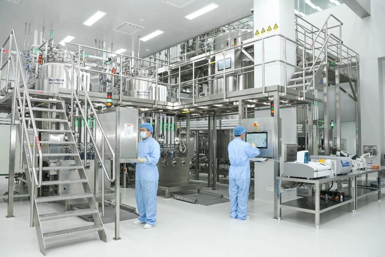
Protein purification buffers constitute an artificial milieu engineered to preserve the structural and functional integrity of target proteins. Far from a simple aqueous salt solution, each buffer is a rigorously defined system comprising a buffering pair, ionic species, and protective excipients. It serves as the medium for column equilibration, sample loading, contaminant washing, and target elution. Upon completion of purification, it remains equally indispensable for resin storage and regeneration. In effect, throughout the entire chromatographic workflow, the purification medium is continuously immersed in buffers of systematically varied composition.
Selection of Protein Purification Buffers
Buffer formulations are dictated by the physicochemical properties of the purification medium and the specific requirements of the process. The following sections outline buffer systems commonly employed with representative chromatographic media at each stage of operation. (The formulations presented herein serve as recommended starting conditions rather than definitive recipes; individual components may be optimized in accordance with the principles set forth above.)
Affinity Chromatography
① His-Tag Affinity
The polyhistidine tag is among the most extensively utilized small affinity tags. Its mechanism of action hinges upon the imidazole moiety of histidine residues, which forms coordinate bonds with immobilized transition metal ions (Ni²⁺, Co²⁺, Cu²⁺, Zn²⁺, etc.), thereby mediating selective retention. Bound protein is subsequently recovered either by competitive elution with elevated imidazole concentrations or by reduction of buffer pH.
Imidazole-Mediated Elution
Equilibration buffer: 50 mM NaH₂PO₄, 300 mM NaCl, 0–10 mM imidazole, pH 8.0
Wash buffer: 50 mM NaH₂PO₄, 300 mM NaCl, 0–50 mM imidazole, pH 8.0
Elution buffer: 50 mM NaH₂PO₄, 300 mM NaCl, 200–300 mM imidazole, pH 8.0
pH-Mediated Elution
Equilibration buffer: 50 mM NaH₂PO₄, 300 mM NaCl, pH 8.0
Wash buffer: 50 mM NaH₂PO₄, 300 mM NaCl, pH 6.3
Elution buffer: 50 mM sodium acetate, 300 mM NaCl, pH 4.5
Purification of inclusion-body-derived proteins: The foregoing formulations are supplemented with 6 M guanidine hydrochloride or 8 M urea to effect solubilization of inclusion bodies and to maintain the target protein in a denatured yet soluble state throughout the chromatographic process.
Notes:
1.Retention is favored under conditions of low imidazole concentration or elevated pH; elution is achieved under conditions of high imidazole concentration or reduced pH.
2.Immobilized metal ions exhibit limited chemical compatibility with chelating and reducing agents such as EDTA and DTT. Where specialized additives are contemplated, reference should be made to the chemical-resistance data supplied in the manufacturer’s product documentation.
② Antibody Affinity
Equilibration buffer: 1× PBS, pH 7.4; alternatively 50 mM Tris-HCl, 0.15 M NaCl, pH 7.5
Wash buffer: 1× PBS, pH 7.4; alternatively 50 mM Tris-HCl, 0.15 M NaCl, pH 7.5
Elution buffer: 0.1 M glycine, pH 3.0; alternatively citrate or acetate buffers, selected on the basis of the stability profile of the target protein
Neutralization buffer: 1 M Tris, pH 8.5–9.0
Note: Eluted antibody fractions require immediate neutralization. Neutralization buffer may be pre-aliquoted into collection vessels; a ratio of 100 μL of neutralization buffer per 1 mL of eluate is recommended.
Ion Exchange Chromatography
Ion exchange chromatography is governed by the principle of retention at low ionic strength and elution at high ionic strength. The selection of buffering species and operating pH is informed by the isoelectric point of the target protein and the class of ion exchanger employed; conversely, the choice of ion exchanger may be predicated upon the protein’s pI and the pH of the matrix in which it is dissolved.
① Interrelationship Among Ion Exchanger Class, Isoelectric Point, and Buffer pH
Anion exchange media: At buffer pH values exceeding the pI, the protein acquires a net negative charge and is retained by the anion exchanger.
Cation exchange media: At buffer pH values below the pI, the protein acquires a net positive charge and is retained by the cation exchanger.
Note: The operating pH is recommended to differ from the protein’s isoelectric point by approximately ±1 pH unit to ensure robust charge-mediated retention.
② Selection of Buffer Components
Equilibration buffer: 10–50 mM buffering salt, devoid of added sodium chloride; conductivity maintained below 10 mS/cm
Wash buffer: equilibration buffer augmented with 100–500 mM NaCl or KCl to displace weakly retained contaminants
Elution buffer: equilibration buffer augmented with elevated concentrations (1–2 M) of NaCl or KCl to effect displacement of strongly retained species
Hydrophobic Interaction Chromatography (HIC)
Hydrophobic interaction chromatography leverages the capacity of high salt concentrations to perturb the protein hydration shell, thereby exposing hydrophobic residues that engage in reversible association with weakly hydrophobic ligands immobilized on the stationary phase. Separation is accomplished by elution under a descending salt gradient. The governing principle is therefore retention at high salt concentration and elution at low salt concentration.
① Equilibration Buffer
Salt selection: Ammonium sulfate is the preferred kosmotropic agent; sodium sulfate or sodium chloride may serve as alternatives. Concentrations typically range from 1.0–1.5 M, and may extend to 2.0 M for proteins with pronounced hydrophobic character.
Buffer system: Phosphate, Tris, or acetate at 20–50 mM, with pH adjusted to accommodate protein stability (commonly 6.0–8.0). Extreme pH conditions conducive to precipitation or denaturation should be avoided.
Note: The ionic strength of the sample must be matched to that of the equilibration buffer to avert protein precipitation arising from osmotic shock upon sample application.
② Elution Buffer
Method 1 (most widely employed): Linear or stepwise reduction of salt concentration to approximately 0 M (e.g., from 1.5 M to 0 M ammonium sulfate).
Method 2: Incorporation of mild organic modifiers (e.g., ethylene glycol, propylene glycol) or low-concentration detergents (for membrane proteins) to attenuate solvent polarity and thereby displace retained protein.
Resin Storage Solutions
The resin storage solution, alternatively termed storage buffer, serves to maintain the purification medium in a hydrated, uniformly dispersed state during periods of inactivity. A bacteriostatic agent is conventionally incorporated to mitigate the risk of microbial proliferation during prolonged storage.
20% ethanol. Ethanol-mediated bacteriostatic protection; appropriate for media bearing small-molecule ligands or protein ligands with demonstrated ethanol tolerance, including ion exchange resins, Ni-chelating resins, and Protein A resins.
2% benzyl alcohol. Associated with higher procurement cost; non-flammable and thus operationally safer; exhibits broad reagent compatibility and a low propensity to induce protein denaturation.
0.05% sodium azide (NaN₃). A potent bacteriostatic agent that exerts no deleterious effect on protein activity; however, its use entails substantial occupational handling and regulatory considerations.
0.1% ProClin 300. A highly efficacious, broad-spectrum bacteriostatic agent with a favorable safety profile; may be employed as a substitute for sodium azide.
Regeneration and Cleaning Solutions
Purification media are generally amenable to repeated use. Nevertheless, with successive cycles, the accumulation of nonspecifically bound material frequently manifests as diminished flow rates and reduced dynamic binding capacity, necessitating intervention by resin cleaning.
Cleaning protocols are medium-specific. The following enumerates representative regeneration cleaning formulations together with their intended applications. The appropriate reagent should be selected with due regard to the chemical and mechanical characteristics of the resin in question.
1–2 M NaCl. Employed for routine cleaning to remove contaminating proteins retained via ionic interactions. Regeneration efficacy is moderate; principally effective against weakly bound species.
6 M guanidine hydrochloride or 8 M urea. Indicated for the resolution of protein aggregation and precipitation. Owing to the propensity of high-salt solutions to crystallize upon concentration, thorough post-cleaning rinsing is imperative to prevent salt deposition.
0.1–1% nonionic detergent. Utilized for the removal of lipophilic contaminants retained via hydrophobic interactions. Detergents are inherently viscous; wash volumes should be increased to ensure quantitative removal.
0.1–1 M sodium hydroxide (NaOH). Employed for the elimination of precipitated material, strongly hydrophobic contaminants, and lipidaceous deposits. Strongly alkaline conditions may compromise affinity ligands; contact time is recommended not to exceed 15 minutes.
Supplementary Additives Commonly Employed
Glycerol. Mitigates protein aggregation and enhances solubility. Recommended concentration: 5–20% (v/v). Elevates solution viscosity; flow rates should be adjusted downward accordingly.
Dithiothreitol (DTT). Preserves sulfhydryl groups against oxidative modification. Recommended concentration: 1–10 mM. Incompatible with Ni-chelating columns (TCEP is the preferred alternative).
Tris(2-carboxyethyl)phosphine (TCEP). A reducing agent of greater potency and stability than DTT. Recommended concentration: 0.5–5 mM. Exerts minimal deleterious effect on metal-chelating columns; generally recommended.
Ethylenediaminetetraacetic acid (EDTA). Chelates divalent heavy metal ions and thereby suppresses metalloprotease activity. Recommended concentration: 0.1–1 mM. Incompatible with metal-chelating affinity columns.
Triton X-100. A nonionic detergent that attenuates nonspecific hydrophobic adsorption. Recommended concentration: 0.1–1%. Must be removed prior to downstream mass spectrometric analysis; incompatible with DTT.
Phenylmethylsulfonyl fluoride (PMSF). An irreversible inhibitor of serine proteases. Recommended concentration: 0.1–1 mM. Exhibits rapid hydrolysis in aqueous media; should be added immediately prior to use.
Formulation of Protein Purification Buffers
Considerations in Buffer Preparation
1. Adherence to a defined operational sequence
Weigh reagents → dissolve in a portion of the final water volume → adjust to the target pH using a calibrated pH meter → q.s. to the final volume. Deviation from this sequence will compromise the accuracy of the final solution volume and the nominal concentrations of constituent reagents.
2. Employment of high-purity water
Buffers should be prepared using deionized or ultrapure water. Where endotoxin-free buffers are required, water certified as endotoxin-free must be used.
3. Temperature control
The pH of certain buffering species is temperature-dependent (notably Tris). Buffers should be prepared and pH-adjusted at the temperature at which subsequent experimentation will be conducted.
4. pH adjustment
① Select acid and base titrants appropriate to the buffering system: for conventional solutions, pH is elevated with NaOH and reduced with HCl; specialized systems require their conjugate acid or base (e.g., acetate buffers should be acidified with acetic acid).
② During titration, add titrant incrementally under continuous agitation and monitor pH progression with a calibrated meter, avoiding iterative over- and under-shooting that may unduly elevate buffer conductivity.
5. Degassing and filtration following preparation
Degassing: Solutions should be degassed by vacuum filtration or sonication for approximately 10 minutes to prevent entrained gas from entering the chromatography column and compromising separation performance.
Filtration: Solutions should be filtered through a 0.45 μm or 0.22 μm membrane to remove particulate matter and insoluble contaminants. A 0.22 μm membrane additionally affords a measure of sterilization, thereby extending the shelf life of the prepared buffer.
6. Preparation of labile reagents immediately prior to use
Certain supplementary components, such as protease inhibitors, are susceptible to degradation upon prolonged storage in aqueous solution. Such reagents should be incorporated into the buffer immediately before use and the resulting solution consumed without undue delay.
Considerations in Buffer Handling and Storage
1.Buffers should be stored in hermetically sealed containers at ambient temperature to prevent reagent precipitation or degradation.
2.Specialized components, such as biotin eluents or peptide eluents, should be aliquoted and stored frozen to avoid repeated freeze-thaw cycling.
3.Buffer should not be aspirated directly from the stock container. The volume required for a given experiment should be dispensed into a separate vessel; any unused remainder should be discarded and never returned to the original container.
4.Buffers exhibiting precipitation, microbial contamination, or other visible deterioration following prolonged storage should be discarded and freshly prepared.